4 0 0 0 OA Spontaneous Middle Meningeal Arteriovenous Fistula Caused by Aneurysm Rupture: A Case Report
- 著者
- Satoshi MIYAMOTO Hisayuki HOSOO Eiichi ISHIKAWA Yuji MATSUMARU
- 出版者
- The Japan Neurosurgical Society
- 雑誌
- NMC Case Report Journal (ISSN:21884226)
- 巻号頁・発行日
- vol.10, pp.81-85, 2023-12-31 (Released:2023-03-24)
- 参考文献数
- 14
Middle meningeal arteriovenous fistula (MMAVF) is a shunt between the middle meningeal artery and the vein surrounding the artery. We report an extremely rare case of spontaneous MMAVF; then, we evaluated the effectiveness of trans-arterial embolization for spontaneous MMAVF and the possible cause of spontaneous MMAVF.A 42-year-old man with tinnitus, a left temporal headache, and pain surrounding the left mandibular joint was diagnosed with MMAVF on digital subtraction angiography. Trans-arterial embolization with detachable coils was conducted, which resulted in a fistula closure and symptoms' diminishment. The cause of MMAVF was thought to be the rupture of the middle meningeal artery aneurysm.A middle meningeal artery aneurysm can be a cause of spontaneous MMAVF, and trans-arterial embolization might be an optimal treatment.
- 著者
- Noriyuki WATANABE Masashi MIZUMOTO Taishi AMANO Hisayuki HOSOO Akinari YAMANO Alexander ZABORONOK Masahide MATSUDA Shingo TAKANO Yuji MATSUMARU Eiichi ISHIKAWA
- 出版者
- The Japan Neurosurgical Society
- 雑誌
- NMC Case Report Journal (ISSN:21884226)
- 巻号頁・発行日
- vol.10, pp.337-342, 2023-12-31 (Released:2023-11-29)
- 参考文献数
- 17
Cavernous sinus hemangioma (CSH) is a rare vascular malformation, arising from the cavernous sinus. Because of its anatomically complex location, a large lesion can cause a variety of symptoms due to cranial nerve compression. A 69-year-old woman with an unsteady gait was admitted to our hospital, and magnetic resonance imaging revealed an extra-axial giant tumor in the cavernous sinus and enlarged ventricles. A radiographic diagnosis of CSH was made. As the risk of surgical removal was considered high, the patient underwent intensity-modulated radiation therapy of 50.4 Gy in 28 fractions. The size of the tumor decreased markedly over time, and the symptoms improved soon after treatment. A 61.8% reduction in tumor size was confirmed immediately after irradiation, and a 75.9% reduction was revealed at a follow-up visit one year later. We reported a case of a giant CSH with hydrocephalus, where tumor shrinkage was confirmed immediately after radiation therapy, and the symptoms of hydrocephalus improved without surgical intervention.
- 著者
- Yoshiro ITO Hisayuki HOSOO Masayuki SATO Aiki MARUSHIMA Mikito HAYAKAWA Yuji MATSUMARU Eiichi ISHIKAWA
- 出版者
- The Japan Neurosurgical Society
- 雑誌
- Neurologia medico-chirurgica (ISSN:04708105)
- 巻号頁・発行日
- pp.2022-0361, (Released:2023-09-23)
- 参考文献数
- 25
In the transsylvian (TS) approach, as characterized by clipping surgery, the presurgical visualization of the superficial middle cerebral vein (SMCV) can help change the surgical approach to ensure safe microsurgery. Nevertheless, identifying preoperatively the venous structures that are involved in this approach is difficult. In this study, we investigated the venous structures that are involved in the TS approach using three-dimensional (3D) rotational venography (3D-RV) and evaluated the effectiveness of this method for presurgical simulation. Patients who underwent 3D-RV between August 2018 and June 2020 were involved in this retrospective study. The 3D-RV and partial maximum intensity projection images with a thickness of 5 mm were computationally reconstructed. The venous structures were subdivided into the following three portions according to the anatomic location: superficial, intermediate, and basal portions. In the superficial portion, predominant frontosylvian veins were observed on 31 (41%) sides, predominant temporosylvian veins on seven (9%) sides, and equivalent fronto- and temporosylvian veins on 28 (37%) sides. The veins in the intermediate (deep middle cerebral and uncal veins) and basal portions (frontobasal bridging veins) emptied into the SMCV on 57 (75%) and 34 (45%) sides, respectively. The 3D-RV images were highly representative of the venous structures observed during microsurgery. In this study, 3D-RV was utilized to capture the details of the venous structures from the superficial to the deep portions. Presurgical simulation of the venous structures that are involved in the TS approach using 3D-RV may increase the safety of microsurgical approaches.
- 著者
- Satoshi Miyamoto Mikito Hayakawa Sho Okune Ryosuke Shintoku Akinari Yamano Takato Hiramine Toshihide Takahashi Hisayuki Hosoo Yoshiro Ito Aiki Marushima Masao Koda Eiichi Ishikawa Yuji Matsumaru
- 出版者
- The Japanese Society of Internal Medicine
- 雑誌
- Internal Medicine (ISSN:09182918)
- 巻号頁・発行日
- pp.1386-22, (Released:2023-06-07)
- 参考文献数
- 9
- 被引用文献数
- 1
Hidden bow hunter's syndrome (HBHS) is a rare disease in which the vertebral artery (VA) occludes in a neutral position but recanalizes in a particular neck position. We herein report an HBHS case and assess its characteristics through a literature review. A 69-year-old man had repeated posterior-circulation infarcts with right VA occlusion. Cerebral angiography showed that the right VA was recanalized only with neck tilt. Decompression of the VA successfully prevented stroke recurrence. HBHS should be considered in patients with posterior circulation infarction with an occluded VA at its lower vertebral level. Diagnosing this syndrome correctly is important for preventing stroke recurrence.
- 著者
- Takao KOISO Yoji KOMATSU Daisuke WATANABE Go IKEDA Hisayuki HOSOO Masayuki SATO Yoshiro ITO Tomoji TAKIGAWA Mikito HAYAKAWA Aiki MARUSHIMA Wataro TSURUTA Noriyuki KATO Kazuya UEMURA Kensuke SUZUKI Akio HYODO Eichi ISHIKAWA Yuji MATSUMARU
- 出版者
- The Japan Neurosurgical Society
- 雑誌
- Neurologia medico-chirurgica (ISSN:04708105)
- 巻号頁・発行日
- pp.2022-0253, (Released:2023-01-05)
- 参考文献数
- 15
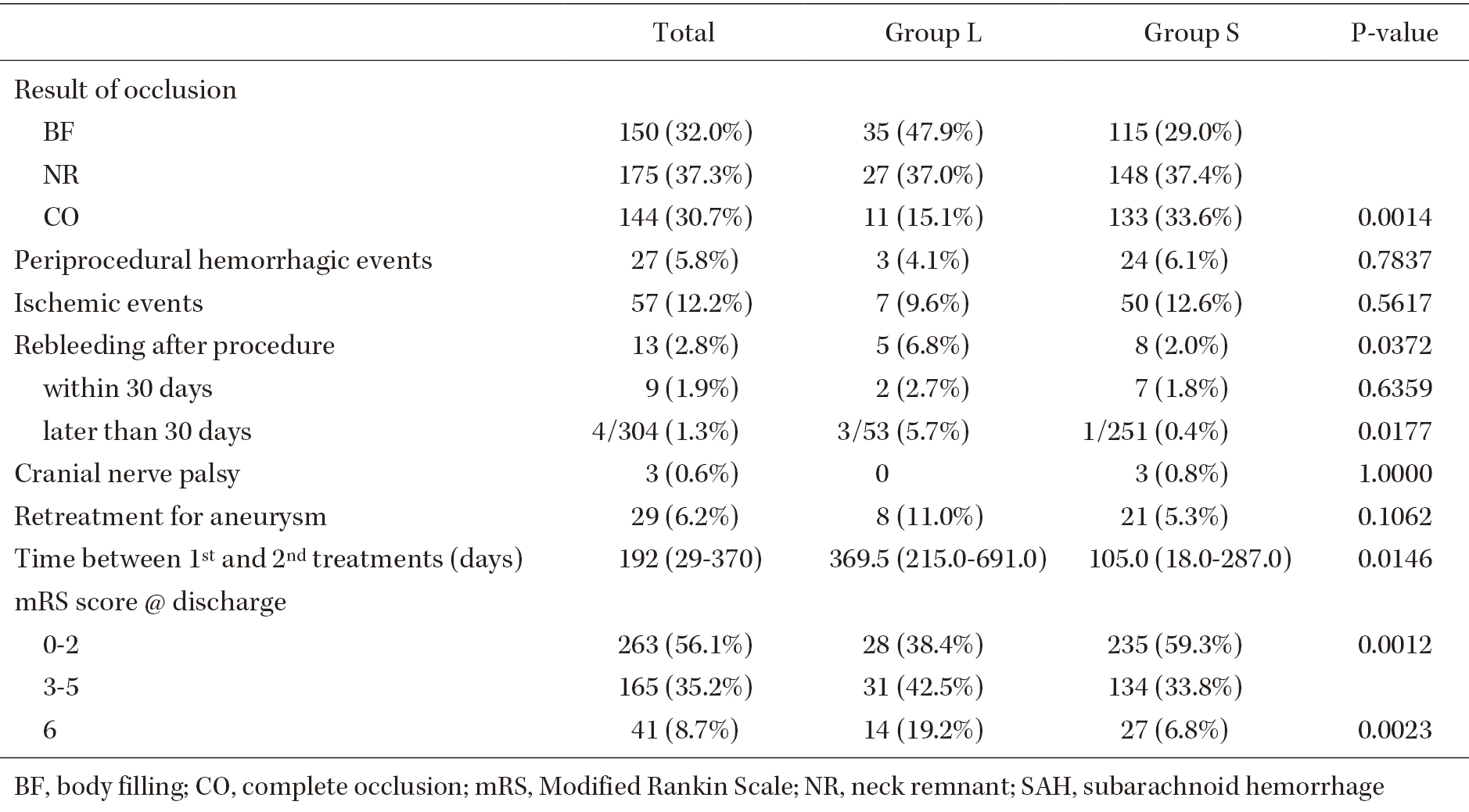
The influence of aneurysm size on the outcomes of endovascular management (EM) for aneurysmal subarachnoid hemorrhages (aSAH) is poorly understood. To evaluate the outcomes of EM for ruptured large cerebral aneurysms, we retrospectively analyzed the medical records of patients with aSAH that were treated with coiling between 2013 and 2020 and compared the differences in outcomes depending on aneurysm size. A total of 469 patients with aSAH were included; 73 patients had aneurysms measuring ≥10 mm in diameter (group L), and 396 had aneurysms measuring <10 mm in diameter (group S). The median age; the percentage of patients that were classified as World Federation of Neurological Surgeons grade 1, 2, or 3; and the frequency of intracerebral hemorrhages differed significantly between group L and group S (p = 0.0105, p = 0.0075, and p = 0.0458, respectively). There were no significant differences in the frequencies of periprocedural hemorrhagic or ischemic events. Conversely, rebleeding after the initial treatment was significantly more common in group L than in group S (6.8% vs. 2.0%; p = 0.0372). The frequency of a modified Rankin Scale score of 0-2 at discharge was significantly lower (p = 0.0012) and the mortality rate was significantly higher (p = 0.0023) in group L than in group S. After propensity-score matching, there were no significant differences in complications and outcomes between the two groups. Rebleeding was more common in large aneurysm cases. However, propensity-score matching indicated that the outcomes of EM for aSAH may not be affected markedly by aneurysm size.


