3 0 0 0 OA Microbleeds Due to Reperfusion Enhance Early Seizures after Carotid Ligation in a Rat Ischemic Model
- 著者
- Takuro SAITO Takeshi MIKAMI Tsukasa HIRANO Hiroshi NAGAHAMA Rei ENATSU Katsuya KOMATSU Satoshi OKAWA Yukinori AKIYAMA Nobuhiro MIKUNI
- 出版者
- The Japan Neurosurgical Society
- 雑誌
- Neurologia medico-chirurgica (ISSN:04708105)
- 巻号頁・発行日
- pp.2022-0372, (Released:2023-04-06)
- 参考文献数
- 35
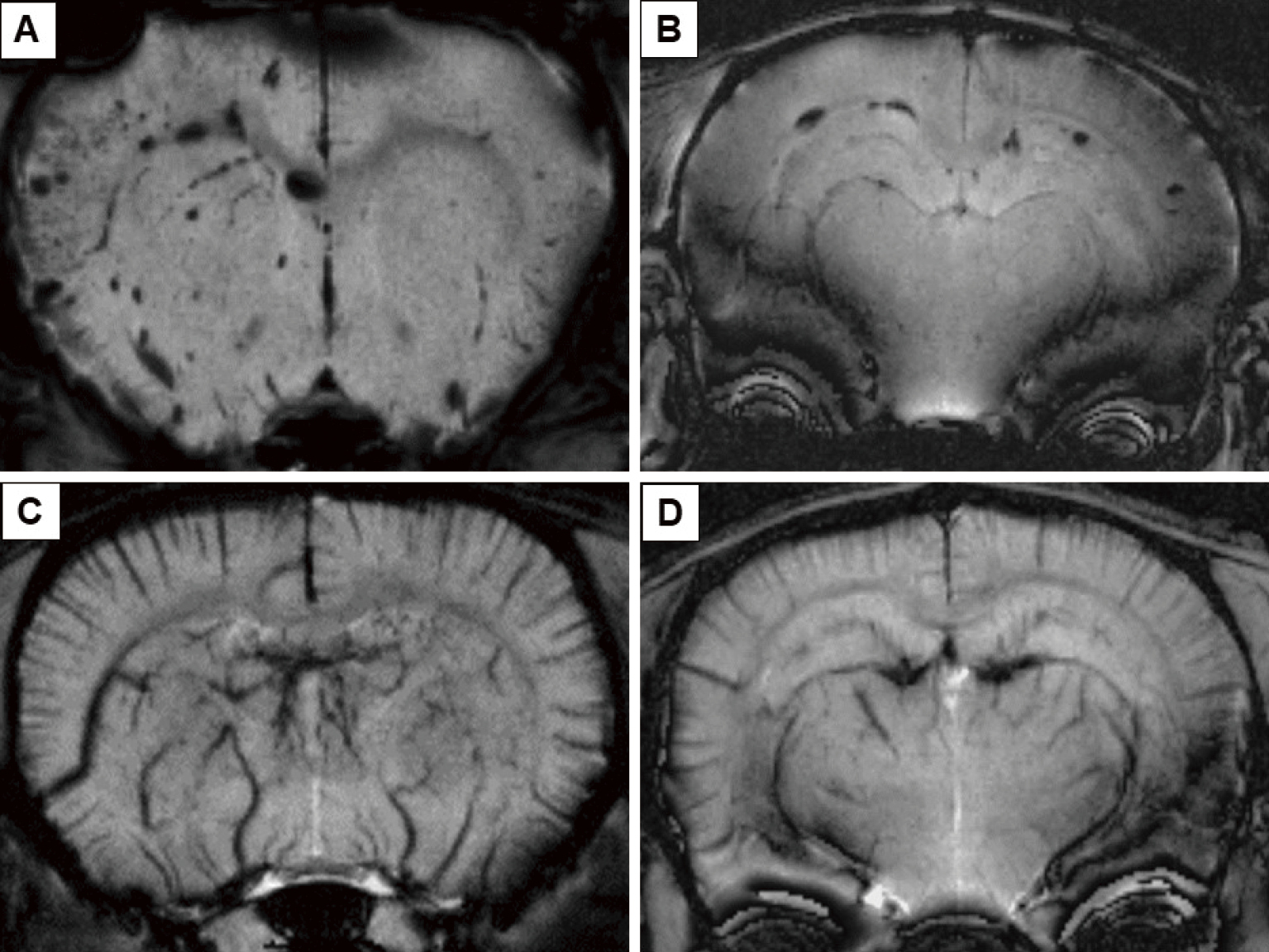
Impaired reperfusion in ischemic brain disease is a condition that we are increasingly confronted with owing to recent advances in reperfusion therapy. In the present study, rat models of reperfusion were investigated to determine the causes of acute seizures using magnetic resonance imaging (MRI) and histopathological specimens. Rat models of bilateral common carotid artery ligation followed by reperfusion and complete occlusion were created. We compared the incidence of seizures, mortality within 24 h, MRI, and magnetic resonance spectroscopy (MRS) to evaluate ischemic or hemorrhagic changes and metabolites in the brain parenchyma. In addition, the histopathological specimens were compared with those observed on MRI. In multivariate analysis, the predictive factors of mortality were seizure (odds ratios (OR), 106.572), reperfusion or occlusion (OR, 0.056), and the apparent diffusion coefficient value of the striatum (OR, 0.396). The predictive factors of a convulsive seizure were reperfusion or occlusion (OR, 0.007) and the number of round-shaped hyposignals (RHS) on susceptibility-weighted imaging (SWI) (OR, 2.072). The incidence of convulsive seizures was significantly correlated with the number of RHS in the reperfusion model. RHS on SWI was confirmed pathologically as microbleeds in the extravasation of the brain parenchyma and was distributed around the hippocampus and cingulum bundle. MRS analysis showed that the N-acetyl aspartate level was significantly lower in the reperfusion group than in the occlusion group. In the reperfusion model, RHS on SWI was a risk factor for convulsive seizures. The location of the RHS also influenced the incidence of convulsive seizures.
1 0 0 0 OA Usefulness of Intraoperative Electrocorticography for the Localization of Epileptogenic Zones
- 著者
- Ryohei CHIBA Rei ENATSU Aya KANNO Tomoaki TAMADA Takuro SAITO Ryota SATO Nobuhiro MIKUNI
- 出版者
- The Japan Neurosurgical Society
- 雑誌
- Neurologia medico-chirurgica (ISSN:04708105)
- 巻号頁・発行日
- pp.2022-0252, (Released:2022-11-25)
- 参考文献数
- 16
- 被引用文献数
- 1
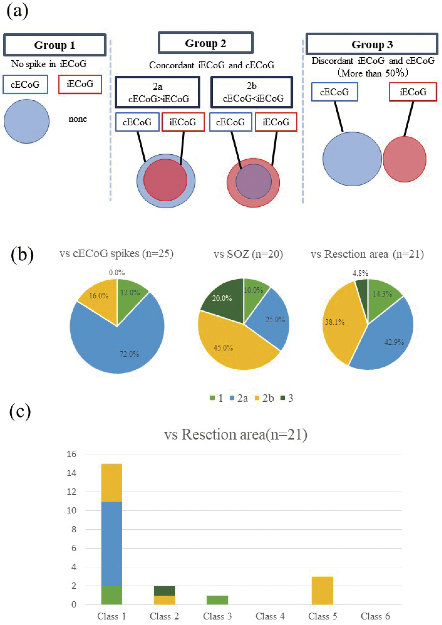
Intraoperative electrocorticography (iECoG) is widely performed to identify irritative zones in the cortex during brain surgery; however, several limitations (e.g., short recording times and the effects of general anesthesia) reduce its effectiveness. The present study aimed to evaluate the utility of iECoG for localizing epileptogenic zones. We compared the results of iECoG and chronic electrocorticography (cECoG) in 25 patients with refractory epilepsy. Subdural electrodes were implanted with iECoG under general anesthesia (2% sevoflurane). cECoG recordings were performed for 3-14 days. The distribution of iECoG spikes was compared with cECoG spike, seizure onset zone, and resection areas. The concordance patterns of each distribution were classified into four patterns: Group 1: No spike in iECoG, Group 2: concordant (2a: iECoG smaller, 2b: iECoG larger, Group 3: discordant >50%). The concordance rate of interictal spikes, seizure onset zones, and resection areas were 88.0% (Group 2a: 72.0%, Group 2b: 16.0%), 70.0% (Group 2a: 25.0%, Group 2b: 45.0%), and 81.0% (Group 2a: 42.9%, Group 2b: 38.1%), respectively. The resection of iECoG spike areas significantly correlated with good surgical outcomes. The indication and limitations of iECoG need to be realized, and the complementary use of iECoG and cECoG may enhance clinical utility.
- 著者
- Seiichiro IMATAKA Rei ENATSU Tsukasa HIRANO Ayaka SASAGAWA Masayasu ARIHARA Tomoyoshi KURIBARA Satoko OCHI Nobuhiro MIKUNI
- 出版者
- The Japan Neurosurgical Society
- 雑誌
- Neurologia medico-chirurgica (ISSN:04708105)
- 巻号頁・発行日
- vol.62, no.5, pp.215-222, 2022-05-15 (Released:2022-05-15)
- 参考文献数
- 24
- 被引用文献数
- 2
The aim of the present study was to evaluate motor area mapping using functional magnetic resonance imaging (fMRI) compared with electrical cortical stimulation (ECS). Motor mapping with fMRI and ECS were retrospectively compared in seven patients with refractory epilepsy in which the primary motor (M1) areas were identified by fMRI and ECS mapping between 2012 and 2019. A right finger tapping task was used for fMRI motor mapping. Blood oxygen level-dependent activation was detected in the left precentral gyrus (PreCG)/postcentral gyrus (PostCG) along the "hand knob" of the central sulcus in all seven patients. Bilateral supplementary motor areas (SMAs) were also activated (n = 6), and the cerebellar hemisphere showed activation on the right side (n = 3) and bilateral side (n = 4). Furthermore, the premotor area (PM) and posterior parietal cortex (PPC) were also activated on the left side (n = 1) and bilateral sides (n = 2). The M1 and sensory area (S1) detected by ECS included fMRI-activated PreCG/PostCG areas with broader extent. This study showed that fMRI motor mapping was locationally well correlated to the activation of M1/S1 by ECS, but the spatial extent was not concordant. In addition, the involvement of SMA, PM/PPC, and the cerebellum in simple voluntary movement was also suggested. Combination analysis of fMRI and ECS motor mapping contributes to precise localization of M1/S1.

