- 著者
- Kenichi Ozawa Susumu Yamamoto Kazuhiko Mase Iwao Matsuda
- 出版者
- The Japan Society of Vacuum and Surface Science
- 雑誌
- e-Journal of Surface Science and Nanotechnology (ISSN:13480391)
- 巻号頁・発行日
- vol.17, pp.130-147, 2019-09-07 (Released:2019-09-07)
- 参考文献数
- 89
- 被引用文献数
- 10
Establishing an accurate view of the photocatalytic mechanism of titanium dioxide (TiO2) has been a challenging task since the discovery of the Honda-Fujishima effect. Despite the great success of catalytic studies in elucidating the chemical and physical aspects of photocatalysis, many questions remain. A surface science approach, which is characterized by the use of atomically well-defined surfaces in precisely controlled environments, is a powerful tool to shed light on the fundamental mechanism, especially the dynamics of photoexcited carriers. In the present contribution, recent progress in photocatalytic research that correlates photocatalytic activity and carrier dynamics on rutile and anatase TiO2 is reviewed. A special focus is placed on the lifetime of photoexcited carriers. We present a method to determine the carrier lifetime; pump-probe time-resolved soft X-ray photoelectron spectroscopy, utilizing an ultraviolet laser as a pump light and a synchrotron radiation as a probe light. The carrier lifetime is found to be linearly correlated with the photocatalytic decomposition/desorption rate of acetic acid adsorbed on single-crystal TiO2 surfaces. The important role of a potential barrier on the TiO2 surface, which influences the carrier lifetime and the photocatalytic activity, is discussed.
- 著者
- Takanori Koitaya Susumu Yamamoto Iwao Matsuda Jun Yoshinobu
- 出版者
- The Japan Society of Vacuum and Surface Science
- 雑誌
- e-Journal of Surface Science and Nanotechnology (ISSN:13480391)
- 巻号頁・発行日
- vol.17, pp.169-178, 2019-11-02 (Released:2019-11-02)
- 参考文献数
- 86
- 被引用文献数
- 4 13
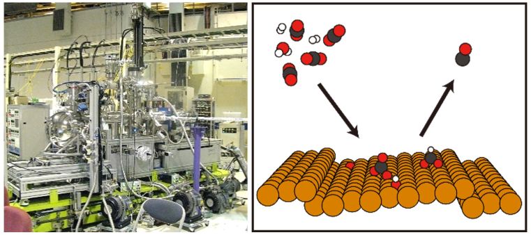
In-situ analysis of heterogeneous catalysts under reaction condition is indispensable to understand reaction mechanisms and nature of active sites. Ambient-pressure X-ray photoelectron spectroscopy (AP-XPS) is one of the powerful methods to investigate chemical states of catalysts and reaction intermediates adsorbed on the surface. In this review, reaction of carbon dioxide on Cu(997) and Zn-deposited Cu(997) surfaces are discussed as an example of surface chemistry of weakly adsorbed molecules, together with a brief overview of recent progress in AP-XPS methods.
- 著者
- Susumu YAMAMOTO Hiroya HASHIZUME Jiro HITOMI Masatsugu SHIGENO Shoichi SAWAGUCHI Haruki ABE Tatsuo USHIKI
- 出版者
- 国際組織細胞学会
- 雑誌
- Archives of Histology and Cytology (ISSN:09149465)
- 巻号頁・発行日
- vol.63, no.2, pp.127-135, 2000 (Released:2005-11-22)
- 参考文献数
- 18
- 被引用文献数
- 29 49
The present study was designed to analyze the subfibrillar structure of corneal and scleral collagen fibrils by scanning electron microscopy (SEM) and atomic force microscopy (AFM). Isolated collagen fibrils of the bovine cornea and sclera were fixed with 1% OsO4, stained with phosphotungstic acid and uranyl acetate, dehydrated in ethanol, critical point-dried, metal-coated, and observed in an in-lens type field emission SEM. Some isolated collagen fibrils were fixed with 1% OsO4, dehydrated, critical point-dried and observed without metal-coating in an AFM. Isolated collagen fibrils treated with acetic acid were also examined by SEM and AFM. SEM and AFM images revealed that corneal and scleral collagen fibrils had periodic transverse grooves and ridges on their surface; the periodicity (i. e., D-periodicity) was about 63 nm in the cornea and about 67 nm in the sclera. Both corneal and scleral collagen fibrils contained subfibrils running helicoidally in a rightward direction to the longitudinal axis of the fibril; the inclination angle was about 15° in the corneal fibrils and 5° in the scleral fibrils. These findings indicate that the different D-periodicity between corneal and scleral fibrils depends on the different inclinations of the subfibrils in each fibril. The present study thus showed that corneal collagen fibrils differ from scleral collagen fibrils not only in diameter but also in substructure.

