- 著者
- Yohei NOUNAKA Shigeyuki TAHARA Kazuma SASAKI Akio MORITA
- 出版者
- The Japan Neurosurgical Society
- 雑誌
- Neurologia medico-chirurgica (ISSN:04708105)
- 巻号頁・発行日
- vol.63, no.1, pp.37-41, 2023-01-15 (Released:2023-01-15)
- 参考文献数
- 6
- 被引用文献数
- 1
Indocyanine green (ICG) is a cyanine dye useful for visualizing blood vessels; it has been developed for endoscopy and is used in skull base surgery. Endoscopy is widely used for hematoma removal after an intracerebral hemorrhage since it is minimally invasive and has a shorter operation time than craniotomy. However, with this technique the surgical field is limited and it is difficult to obtain an adequate orientation; thus, it is challenging to locate the bleeding point, and postoperative rebleeding has been reported. We performed intraoperative ICG near-infrared fluorescence imaging to locate the bleeding point. This purpose of this study was to evaluate the usefulness of ICG angiography during endoscopic hematoma removal in two patients, using two endoscope types and comparing their visualization of perforating branches during the procedure. ICG angiography was performed in two different cases of putaminal hemorrhage, using the SPIES NIR/ICG-System and IMAGE1 S Rubina (both KARL STORZ, Tuttlingen, Germany) at the intraoperative bleeding site. The intraoperative use of ICG allowed the clear visualization of the perforating branches and real-time confirmation of active bleeding. We could also distinguish an old hematoma from the active bleeding point. The IMAGE1 S Rubina has adequate brightness for contrast enhancement, allowing surgical manipulation simultaneously to the enhancement phase.ICG fluorescence angiography is useful to identify the damaged vessel and perform hemostasis. We expect other similar devices to be developed in the future, accompanied by flexible and thin rigid endoscopes.
- 著者
- Yasuo MURAI Fumihiro MATANO Koshiro ISAYAMA Yohei NOUNAKA Akio MORITA
- 出版者
- The Japan Neurosurgical Society
- 雑誌
- Neurologia medico-chirurgica (ISSN:04708105)
- 巻号頁・発行日
- vol.62, no.11, pp.530-534, 2022-11-15 (Released:2022-11-15)
- 参考文献数
- 25
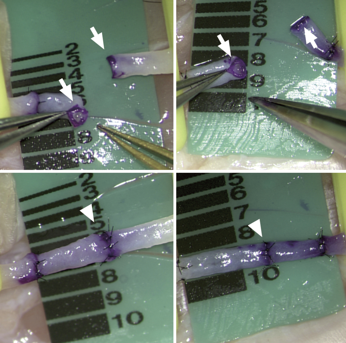
Crystal violet (CV) ink has been used as a skin marker worldwide. It has been reported to be useful for vessel wall visualization of microvascular anastomoses. Contrastingly, it has been found to be carcinogenic and inhibit migration and proliferation of venous cells. In some countries, its use in the medical field has been restricted. Therefore, it is necessary to consider alternatives to CV. In this present study, we compared the time required for the anastomosis of a 0.8-1 mm diameter vessel in the chicken wrist artery using CV and a CV-free dye (ethyl violet; EV). The surgeon, microscope, and anastomosis microsurgical tools were standardized for comparison. CV and EV were changed for each anastomosis. The same surgeon performed 30 anastomoses using each dye. No visually obvious differences were noted in the vascular transections with CV and EV. As per the results, no statistically significant difference was observed in the time required for anastomosis using CV and EV. EV conforming to California Proposition 65 may be an effective alternative to CV for vascular visualization of microvascular anastomoses. However, further studies on the effectiveness of the EV in clinical cases are needed.

