- 著者
- Kento TAKAHARA Tomoru MIWA Takashi IWAMA Masahiro TODA
- 出版者
- The Japan Neurosurgical Society
- 雑誌
- NMC Case Report Journal (ISSN:21884226)
- 巻号頁・発行日
- vol.10, pp.185-189, 2023-12-31 (Released:2023-06-26)
- 参考文献数
- 27
The occipital transtentorial approach (OTA), which is often applied for superior cerebellar lesions, has an inevitable risk of homonymous hemianopsia due to the retraction of the occipital lobe. The endoscopic approach provides increased visibility of the surgical field due to the wide-angled panoramic view and is minimally invasive in approaching deep brain lesions compared to the conventional microscopic approach. However, little is known regarding endoscopic OTA for the removal of cerebellar lesions. We experienced a case of a hemangioblastoma in the paramedian superior surface of the cerebellum that was successfully treated with endoscopic OTA combined with gravity retraction while avoiding postoperative visual dysfunction.A 48-year-old woman was diagnosed with a hemangioblastoma in the superior surface of the cerebellum. She underwent tumor removal with endoscopic OTA combined with gravity retraction of the occipital lobe instead of using brain retractors. The narrower space was sufficient for surgical manipulation with a panoramic view obtained by endoscopy. The simultaneous observation of the lesion with both an endoscope and a microscope revealed the superiority of infratentorial visualization with an endoscope. Gross total removal was achieved with no postoperative complications, including visual dysfunction.Endoscopic OTA may reduce the risk of postoperative visual dysfunction because of its minimally invasive nature, which is enhanced when combined with gravity retraction. Additionally, the panoramic view of the endoscope allows favorable visualization of an infratentorial lesion, which is otherwise hidden partly by the tentorium. The use of endoscopy is compatible with OTA, and endoscopic OTA could be an option for superior cerebellar lesions for avoiding visual dysfunction.
- 著者
- Kento TAKAHARA Satoshi TAKAHASHI Utaro HINO Takashi HORIGUCHI Masahiro TODA
- 出版者
- The Japan Neurosurgical Society
- 雑誌
- NMC Case Report Journal (ISSN:21884226)
- 巻号頁・発行日
- vol.9, pp.377-382, 2022-12-31 (Released:2022-11-09)
- 参考文献数
- 19
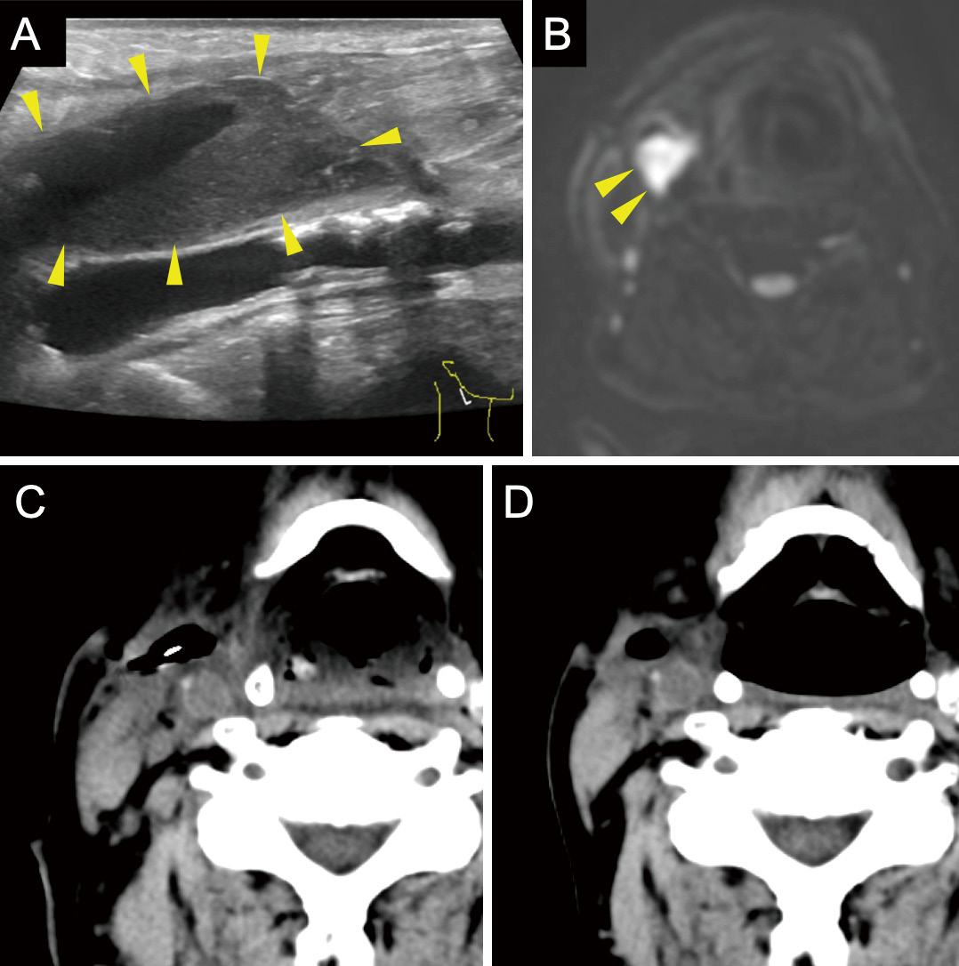
Carotid endarterectomy (CEA) and carotid artery stenting (CAS) for internal carotid artery (ICA) stenosis have specific risks. Therefore, the accurate evaluation and management of each risk factor are important, especially for patients who are at high risk for both CEA and CAS.We report the case of a 77-year-old man with right ICA stenosis that progressed despite optimal medical treatment. In addition, he had several risk factors for both CEA and CAS, including previous cervical radiation therapy, contralateral ICA occlusion, chronic kidney insufficiency, and severe aortic valve stenosis. CEA was performed with priority given to aortic valve stenosis without complications, and the patient was discharged 10 days postoperatively, without neurological sequelae. However, a pericarotid cervical abscess was detected by carotid echo, computed tomography (CT), and magnetic resonance imaging (MRI) 1 month after CEA that required surgical drainage. The infection was thought to be odontogenic because the pathogen was identified as normal oral bacterial flora, and a wound infection was not apparent. Teeth extraction and abscess drainage, in combination with antibiotic therapy, successfully cured the infection without additional complications.Odontogenic cervical abscesses after CEA can occur, especially if the patient is at risk of infection. Therefore, both preoperative and postoperative dental evaluation and management are recommended. As in this case, a cervical abscess can occur without wound infection, and the abscess diagnosis is sometimes difficult from wound inspection alone. Cervical echocardiogram and CT were useful for detecting fluid collection, whereas MRI was useful for qualitatively evaluating the lesion.

