- 著者
- Mao YOKOTA Koji OSUKA Yusuke OHMICHI Mika OHMICHI Chiharu SUZUKI Masahiro AOYAMA Kenichiro IWAMI Satoru HONMA Shigeru MIYACHI
- 出版者
- The Japan Neurosurgical Society
- 雑誌
- Neurologia medico-chirurgica (ISSN:04708105)
- 巻号頁・発行日
- pp.2023-0079, (Released:2023-11-30)
- 参考文献数
- 27
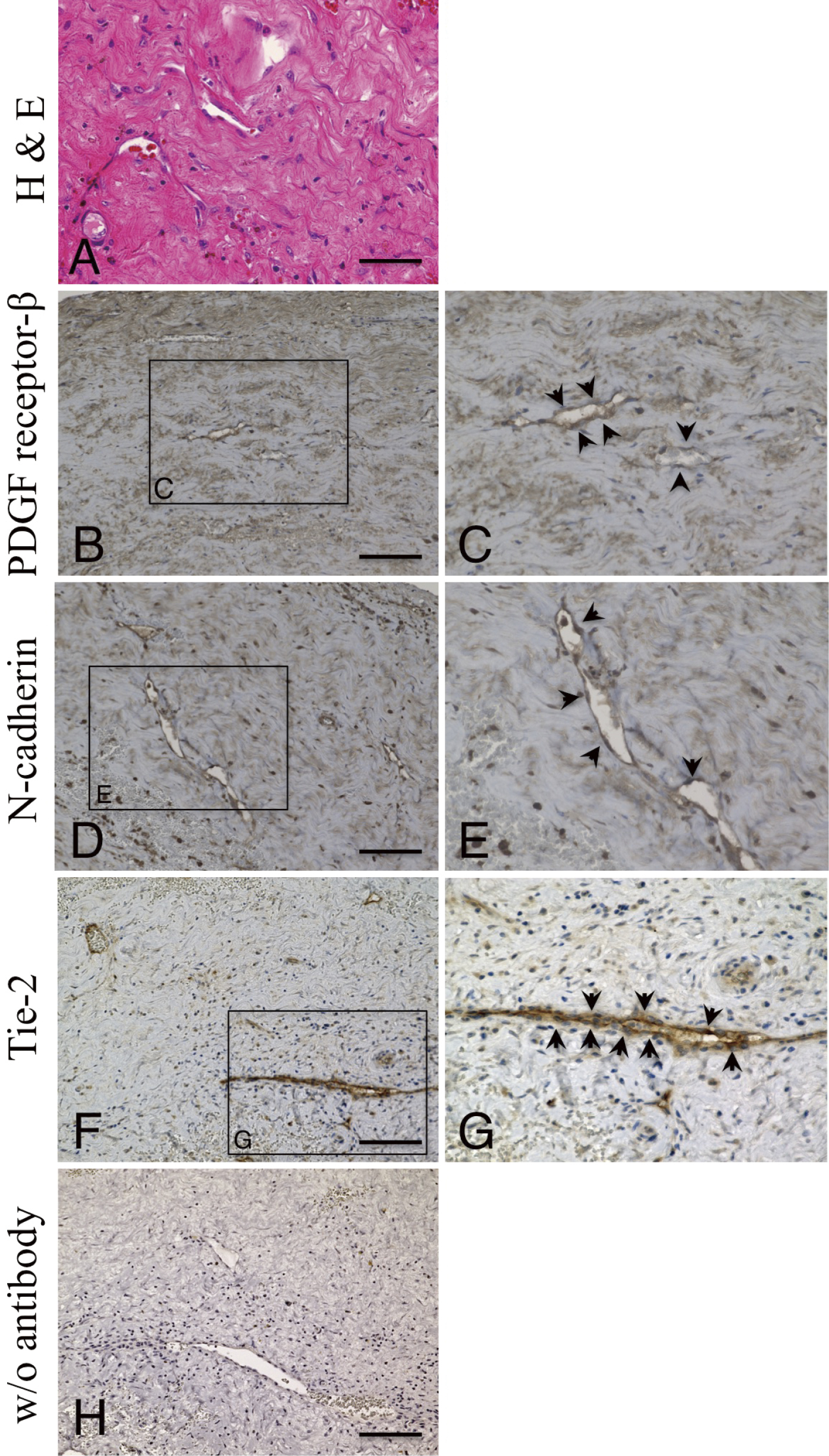
Angiogenesis is one of the growth mechanisms of chronic subdural hematoma (CSDH). Pericytes have been implicated in the capillary sprouting during angiogenesis and are involved in brain ischemia and diabetic retinopathy. This study examined the pericyte expressions in CSDH outer membranes obtained during trepanation surgery. Eight samples of CSDH outer membranes and 35 samples of CSDH fluid were included. NG2, N-cadherin, VE-cadherin, Tie-2, endothelial nitric oxide synthase (eNOS), platelet-derived growth factor (PDGF) receptor-β (PDGFR-β), a well-known marker of pericytes, phosphorylated PDGFR-β at Tyr751, and β-actin expressions, were examined using western blot analysis. PDGFR-β, N-cadherin, and Tie-2 expression levels were also examined using immunohistochemistry. The concentrations of PDGF-BB in CSDH fluid samples were measured using enzyme-linked immunosorbent assay kits. NG2, N-cadherin, VE-cadherin, Tie-2, eNOS, PDGFR-β, and eNOS expressions in CSDH outer membranes were confirmed in all cases. Furthermore, phosphorylated PDGFR-β at Tyr751 was also detected. In addition, PDGFR-β, N-cadherin, and Tie-2 expressions were localized to the endothelial cells of the vessels within CSDH outer membranes by immunohistochemistry. The concentration of PDGF-BB in CSDH fluids was significantly higher than that in cerebrospinal fluid. These findings indicate that PDGF activates pericytes in the microvessels of CSDH outer membranes and suggest that pericytes are crucial in CSDH angiogenesis through the PDGF/PDGFR-β signaling pathway.
- 著者
- Masazumi FUJII Satoshi MAESAWA Sumio ISHIAI Kenichiro IWAMI Miyako FUTAMURA Kiyoshi SAITO
- 出版者
- The Japan Neurosurgical Society
- 雑誌
- Neurologia medico-chirurgica (ISSN:04708105)
- 巻号頁・発行日
- vol.56, no.7, pp.379-386, 2016 (Released:2016-07-15)
- 参考文献数
- 57
- 被引用文献数
- 24 41
The neural basis of language had been considered as a simple model consisting of the Broca’s area, the Wernicke’s area, and the arcuate fasciculus (AF) connecting the above two cortical areas. However, it has grown to a larger and more complex model based upon recent advancements in neuroscience such as precise imaging studies of aphasic patients, diffusion tensor imaging studies, functional magnetic resonance imaging studies, and electrophysiological studies with cortical and subcortical stimulation during awake surgery. In the present model, language is considered to be processed through two distinct pathways, the dorsal stream and the ventral stream. The core of the dorsal stream is the superior longitudinal fasciculus/AF, which is mainly associated with phonological processing. On the other hand, semantic processing is done mainly with the ventral stream consisting of the inferior fronto-occipital fasciculus and the intratemporal networks. The frontal aslant tract has recently been named the deep frontal tract connecting the supplementary motor area and the Broca’s area and it plays an important role in driving and initiating speech. It is necessary for every neurosurgeon to have basic knowledge of the neural basis of language. This knowledge is essential to plan safer surgery and preserve the above neural structures during surgery.
