- 著者
- Babita Shashni Shinya Ariyasu Reisa Takeda Toshihiro Suzuki Shota Shiina Kazunori Akimoto Takuto Maeda Naoyuki Aikawa Ryo Abe Tomohiro Osaki Norihiko Itoh Shin Aoki
- 出版者
- The Pharmaceutical Society of Japan
- 雑誌
- Biological and Pharmaceutical Bulletin (ISSN:09186158)
- 巻号頁・発行日
- vol.41, no.4, pp.487-503, 2018-04-01 (Released:2018-04-01)
- 参考文献数
- 45
- 被引用文献数
- 34 65
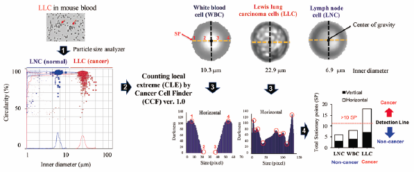
Detection of anomalous cells such as cancer cells from normal blood cells has the potential to contribute greatly to cancer diagnosis and therapy. Conventional methods for the detection of cancer cells are usually tedious and cumbersome. Herein, we report on the use of a particle size analyzer for the convenient size-based differentiation of cancer cells from normal cells. Measurements made using a particle size analyzer revealed that size parameters for cancer cells are significantly greater (e.g., inner diameter and width) than the corresponding values for normal cells (white blood cells (WBC), lymphocytes and splenocytes), with no significant difference in shape parameters (e.g., circularity and convexity). The inner diameter of many cancer cell lines is greater than 10 µm, in contrast to normal cells. For the detection of WBC having similar size to that of cancer cells, we developed a PC software “Cancer Cell Finder” that differentiates them from cancer cells based on brightness stationary points on a cell surface. Furthermore, the aforementioned method was validated for cancer cell/clusters detection in spiked mouse blood samples (a B16 melanoma mouse xenograft model) and circulating tumor cell cluster-like particles in the cat and dog (diagnosed with cancer) blood samples. These results provide insights into the possible applicability of the use of a particle size analyzer in conjunction with PC software for the convenient detection of cancer cells in experimental and clinical samples for theranostics.
