6 0 0 0 OA 講義「新旧古典で解きなおす現代アメリカ」を終えて
- 著者
- 石川 敬史 大泉 惟 鈴木 俊弘 関口 洋平 松原 宏之 イシカワ タカフミ オオイズミ ユイ スズキ トシヒロ セキグチ ヨウヘイ マツバラ ヒロユキ Takafumi Ishikawa Yui Oizumi Toshihiro Suzuki Yohei Sekiguchi Hiroyuki Matsubara
- 雑誌
- 立教アメリカン・スタディーズ = Rikkyo American Studies
- 巻号頁・発行日
- vol.43, pp.115-138, 2021
- 著者
- Babita Shashni Shinya Ariyasu Reisa Takeda Toshihiro Suzuki Shota Shiina Kazunori Akimoto Takuto Maeda Naoyuki Aikawa Ryo Abe Tomohiro Osaki Norihiko Itoh Shin Aoki
- 出版者
- The Pharmaceutical Society of Japan
- 雑誌
- Biological and Pharmaceutical Bulletin (ISSN:09186158)
- 巻号頁・発行日
- vol.41, no.4, pp.487-503, 2018-04-01 (Released:2018-04-01)
- 参考文献数
- 45
- 被引用文献数
- 34 65
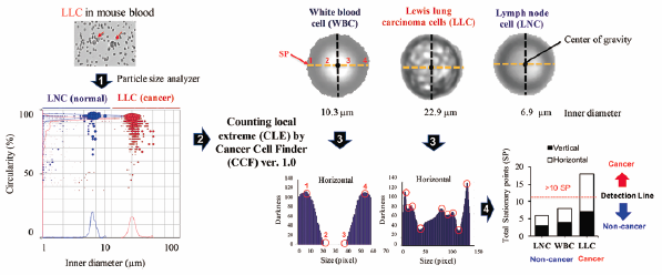
Detection of anomalous cells such as cancer cells from normal blood cells has the potential to contribute greatly to cancer diagnosis and therapy. Conventional methods for the detection of cancer cells are usually tedious and cumbersome. Herein, we report on the use of a particle size analyzer for the convenient size-based differentiation of cancer cells from normal cells. Measurements made using a particle size analyzer revealed that size parameters for cancer cells are significantly greater (e.g., inner diameter and width) than the corresponding values for normal cells (white blood cells (WBC), lymphocytes and splenocytes), with no significant difference in shape parameters (e.g., circularity and convexity). The inner diameter of many cancer cell lines is greater than 10 µm, in contrast to normal cells. For the detection of WBC having similar size to that of cancer cells, we developed a PC software “Cancer Cell Finder” that differentiates them from cancer cells based on brightness stationary points on a cell surface. Furthermore, the aforementioned method was validated for cancer cell/clusters detection in spiked mouse blood samples (a B16 melanoma mouse xenograft model) and circulating tumor cell cluster-like particles in the cat and dog (diagnosed with cancer) blood samples. These results provide insights into the possible applicability of the use of a particle size analyzer in conjunction with PC software for the convenient detection of cancer cells in experimental and clinical samples for theranostics.
- 著者
- Takafumi Hirata Shuji Yamashita Mirai Ishida Toshihiro Suzuki
- 出版者
- The Mass Spectrometry Society of Japan
- 雑誌
- Mass Spectrometry (ISSN:2187137X)
- 巻号頁・発行日
- vol.9, no.1, pp.A0085, 2020-06-10 (Released:2020-06-12)
- 参考文献数
- 45
- 被引用文献数
- 12
We measured the Re/Os (185Re/188Os) and 187Os/188Os ratios from nanoparticles (NPs) using a multiple collector-inductively coupled plasma-mass spectrometer equipped with high-time resolution ion counters (HTR-MC-ICP-MS). Using the HTR-MC-ICP-MS system developed in this study, the simultaneous data acquisition of four isotopes was possible with a time resolution of up to 10 μs. This permits the quantitative analysis of four isotopes to be carried out from transient signals (e.g., <0.6 ms) emanating from the NPs. Iridium–Osmium NPs were produced from a naturally occurring Ir–Os alloy (ruthenosmiridium from Hokkaido, Japan; osmiridium from British Columbia, Canada; iridosmine from the Urals region of Russia) through a laser ablation technique, and the resulting nanoparticles were collected by bubbling water through a suspension. The 187Os/188Os ratios for individual NPs varied significantly, mainly due to the counting statistics of the 187Os and 188Os signals. Despite the large variation in the measured ratios, the resulting 187Os/188Os ratios for three Ir–Os bearing minerals, were 0.121±0.013 for Hokkaido, 0.110±0.012 for British Columbia, and 0.122±0.020 for the Urals, and these values were in agreement with the ratios obtained by the conventional laser ablation-MC-ICP-MS technique. The data obtained here provides a clear demonstration that the HTR-MC-ICP-MS technique can become a powerful tool for monitoring elemental and isotope ratios from NPs of multiple components.
- 著者
- Kiyomi Kohinata Kunihito Matsumoto Toshihiro Suzuki Mari Tsunoda Yusuke Hayashi Masao Araki Koji Hashimoto Kazuya Honda
- 出版者
- 日本大学歯学部
- 雑誌
- Journal of Oral Science (ISSN:13434934)
- 巻号頁・発行日
- vol.58, no.1, pp.29-34, 2016 (Released:2016-03-26)
- 参考文献数
- 22
- 被引用文献数
- 5 1
As part of our ongoing investigation of risk and predictive factors associated with temporomandibular disorders, we used magnetic resonance imaging (MRI) to identify risk factors for sideways disk displacement of the temporomandibular joint in 26 patients with MRI-confirmed unilateral pure sideways disk displacement (medial or lateral disk displacement) and normal positioning of the contralateral temporomandibular joint. Coronal morphologic harmonization between the condyle and fossa, angle between the axis of the ramus and condyle, and angle between the lateral pterygoid muscle (LPM) and condyle were evaluated. Only angle of the LPM related to the condyle was significantly correlated with mediolateral disk position; the angles of joints with medial, normal, and lateral disk positions were 70.2°, 66.7°, and 60.1°, respectively. These results suggest that a greater angle of the inferior head of the LPM to the axis of the condyle on axial MRI images may cause medial disk displacement, while a smaller angle may result in lateral disk displacement. (J Oral Sci 58, 29-34, 2016)
- 著者
- Kunihiko Sawada Toshihiko Amemiya Shigenori Hirai Yusuke Hayashi Toshihiro Suzuki Masahiko Honda Johnny Sisounthone Kunihito Matsumoto Kazuya Honda
- 出版者
- Nihon University School of Dentistry
- 雑誌
- Journal of Oral Science (ISSN:13434934)
- 巻号頁・発行日
- vol.60, no.1, pp.137-141, 2018 (Released:2018-03-24)
- 参考文献数
- 23
- 被引用文献数
- 5
We compared the diagnostic reliability of 3.0-T magnetic resonance imaging (MRI) for detection of osseous abnormalities of the temporomandibular joint (TMJ) with that of the gold standard, cone-beam computed tomography (CBCT). Fifty-six TMJs were imaged with CBCT and MRI, and images of condyles and fossae were independently assessed for the presence of osseous abnormalities. The accuracy, sensitivity, and specificity of 3.0-T MRI were 0.88, 1.0, and 0.73, respectively, in condyle evaluation and 0.91, 0.75, and 0.95 in fossa evaluation. The McNemar test showed no significant difference (P > 0.05) between MRI and CBCT in the evaluation of osseous abnormalities in condyles and fossae. The present results indicate that 3.0-T MRI is equal to CBCT in the diagnostic evaluation of osseous abnormalities of the mandibular condyle.
1 0 0 0 OA iQuant2: Software for Rapid and Quantitative Imaging Using Laser Ablation-ICP Mass Spectrometry
- 著者
- Toshihiro Suzuki Shuhei Sakata Yoshiki Makino Hideyuki Obayashi Seiya Ohara Kentaro Hattori Takafumi Hirata
- 出版者
- The Mass Spectrometry Society of Japan
- 雑誌
- Mass Spectrometry (ISSN:2187137X)
- 巻号頁・発行日
- vol.7, no.1, pp.A0065-A0065, 2018-02-23 (Released:2018-03-01)
- 参考文献数
- 14
- 被引用文献数
- 37
We report on the development of a software program named iQuant2 which creates visual images from two-dimensional signal intensity data obtained by a laser ablation-ICP-mass spectrometry (LA-ICPMS) technique. Time-resolved signal intensity profiles can be converted to position resolved signal intensity data based on the rastering rate (μm s−1) of the laser ablation. Background signal intensities obtained without laser ablation (gas blank) are used as the background, and all of the blank-subtracted intensity data can be used for the imaging analysis. With this software, deformation of the created image can be corrected visually on a PC screen. The line profile analysis between the user-selected points can be observed using the iQuant2 software. To accomplish this, data points on the profile line were automatically calculated based on the interpolation between the analysis points. The resulting imaging data can be exported and stored as JPEG, BMP or PNG formats for further processing. Moreover, a semi-quantitative analysis can be made based on the coupling of the external correction of the RSF (relative sensitivity factor) using NIST SRM610 with normalization of the corrected signal intensity data being 100%. The calculated abundance data for major elements are in reasonable agreement with the values obtained by electron probe micro analyzer (EPMA). With the software developed in this study, both the rapid imaging and semi-quantitative determinations can be made.
- 著者
- Kunihito Matsumoto Souksavanh Vongsa Ichiro Nakajima Ken-ichiro Ejima Kiyomi Kohinata Toshihiro Suzuki Shigeharu Hosono Hirofumi Aboshi Fumiyuki Kuwata Kichibee Otsuka
- 出版者
- 日本大学歯学部
- 雑誌
- Journal of Oral Science (ISSN:13434934)
- 巻号頁・発行日
- vol.57, no.3, pp.235-239, 2015 (Released:2015-09-15)
- 参考文献数
- 12
- 被引用文献数
- 2 4
As part of quality assessment of a teleradiology program we evaluated the validity of patient information received, the quality of panoramic radiography imaging in Laos, and the ability of a Laotian radiologist to detect temporomandibular joint abnormalities. The amount of patient information gathered from 2,021 scans of panoramic radiographs was evaluated by triage before image diagnosis. Among the radiographs from 2,021 patients, primary triage indicated that there was insufficient information for 794 (39.3%) patients. Secondary triage to assess imaging failure included 1,227 radiographs, four of which were excluded from imaging diagnosis because of unacceptable image flaws. In total, 2,446 joints from 1,223 radiographs were evaluated for temporomandibular joint abnormalities in order to compare the image interpretation abilities of Laotian and Japanese radiologists. The kappa coefficient was 0.836 (P < 0.01) for the agreement between the two observers in detecting temporomandibular joint abnormalities on radiographs. We conclude that additional efforts are needed in order to overcome the challenges of maintaining quality in imaging techniques and diagnoses in Laos. (J Oral Sci 57, 235-239, 2015)
