- 著者
- Kengo KISHIDA Daisuke MARUYAMA Saki KOTANI Nobukuni MURAKAMI Naoya HASHIMOTO
- 出版者
- The Japan Neurosurgical Society
- 雑誌
- Neurologia medico-chirurgica (ISSN:04708105)
- 巻号頁・発行日
- pp.2023-0043, (Released:2023-11-08)
- 参考文献数
- 41
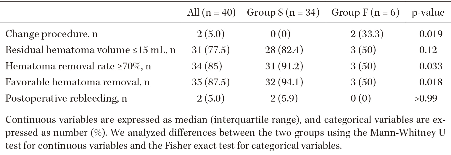
Studies regarding hematoma stiffness and removal difficulty are scarce. This study explored the association between hematoma stiffness and surgical results of endoscopic hematoma removal for intracerebral hemorrhage. It also aimed to clarify factors associated with hematoma stiffness. We classified intracerebral hematoma as either soft or firm stiffness by retrospectively evaluating operative videos by two neurosurgeons. The interobserver reliability of the classification was assessed by calculating the κ values. We investigated the relationship between hematoma stiffness and surgical results. Favorable hematoma removal (FHR) was defined as a residual hematoma volume of ≤15 mL or removal rate of ≥70%. Furthermore, we compared the background characteristics, imaging findings, and laboratory data between the two groups. Forty patients were included in this study. The mean baseline hematoma volume was 69.9 mL (range, 41.3-97.6 mL). FHR was accomplished in 35 cases (87.5%). Thirty-four patients (85%) were in the soft hematoma group (group S). Six patients (15%) were in the firm hematoma group (group F). Classification of hematoma stiffness demonstrated an excellent degree of interobserver agreement (κ score = 0.91). Patients in group S had a high FHR rate (p = 0.018) and short endoscopic procedure times (p = 0.00034). The island sign was present in group S (p = 0.030). Patients in group F had significantly high fibrinogen levels (p = 0.049) and low serum total calcium (p = 0.032), hemoglobin (p = 0.041), and hematocrit (p = 0.011) levels. Hematoma stiffness during endoscopic surgery for intracerebral hemorrhage correlates with surgical results, including the endoscopic procedure time and accomplishing rate of FHR.
- 著者
- Mina Ohtsu Daisuke Kurihara Yoshikatsu Sato Takuya Suzaki Masayoshi Kawaguchi Daisuke Maruyama Tetsuya Higashiyama
- 出版者
- 日本メンデル協会、国際細胞学会
- 雑誌
- CYTOLOGIA (ISSN:00114545)
- 巻号頁・発行日
- vol.82, no.3, pp.251-259, 2017-06-25 (Released:2017-09-06)
- 参考文献数
- 35
Nematode infection of plant roots is a paradigm of host–parasite interactions. Although nematodes can be labeled with fluorescent dyes, migration of the worms into the deep regions of host roots makes them difficult to track. Here we report the use of two fluorescent dyes, FM4-64 and SYBR green I, to intensely label the soybean cyst nematode (SCN) Heterodera glycines for one week in host plants. Continuous monitoring of the labeled SCN juveniles was achieved with two-photon microscopy. Additionally, we developed a transient transformation system consisting of the non-model leguminous plant (fabaceous) roots, Astragalus sinicus and Agrobacterium rhizogenes to observe the cellular structures of the plant during SCN infection. By the combined use of fluorescent dyes and two-photon microscopy, clear images of infecting SCNs were obtained even in deep regions of A. sinicus roots. The fluorescent labeling described herein can also be used in detailed monitoring of the infection processes of other non-model nematodes, as well as the associated morphological changes in the host plant roots.
