- 著者
- 河内 修司 蔵原 晃一 川崎 啓祐 大城 由美 高橋 郁雄 八板 弘樹 岡本 康治 永田 豊 澤野 美由紀 吉岡 翼 渕上 忠彦
- 出版者
- 医学書院
- 巻号頁・発行日
- pp.1318-1325, 2014-08-25
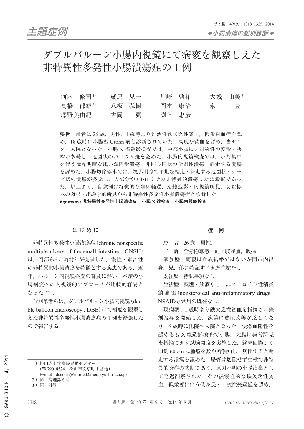
要旨 患者は26歳,男性.1歳時より難治性鉄欠乏性貧血,低蛋白血症を認め,18歳時に小腸型Crohn病と診断されていた.高度な貧血を認め,当センター入院となった.小腸X線造影検査では,中部小腸に非対称性の変形・狭窄が多発し,地図状のバリウム斑を認めた.小腸内視鏡検査では,ひだ集中を伴う境界明瞭な浅い類円形潰瘍,非同心円状の全周性潰瘍,斜走する潰瘍を認めた.小腸切除標本では,境界明瞭で平坦な輪走・斜走する地図状・テープ状の潰瘍が多発し,大部分がUl-IIまでの非特異的潰瘍または瘢痕であった.以上より,自験例は特徴的な臨床経過,X線造影・内視鏡所見,切除標本の肉眼・組織学的所見から非特異性多発性小腸潰瘍症と診断した.
- 著者
- 高橋 郁雄 佐保 春芳
- 出版者
- 北方森林学会
- 雑誌
- 日本林學會北海道支部講演集
- 巻号頁・発行日
- vol.20, pp.181-186, 1972
The larch canker was reported in the Tokyo University Forest in Hokkaido in 1969. Its pathogen, Encoeliorsis laricina (E_<TTL>.) G_<ROVES> (Imperfect state : Brunchorstia laricina E_<TTL>.). was cultured on a potato-galactose-ager medium and fungus colonies on this medium were used for the inoculation experiments. The inoculations were made on Feb. 17, 1970 and March, 16, 1970 on the potted larch seedlings in the green house, quite earlier than the natural condition. The methods of inoculation was as follows. Each main stem and side branch of seedlings were sterilized with 80% alcohol and washed with water, and a peeling abrasion (4mm x 4mm) was made with a sterilized knife on the bark. The pieces of the mycelial colony were placed at the injured part. After that, the plants were placed in a moist chamber and held at the relative humidity of 80-100% and at 15℃ temperature for two weeks. Then, they were placed in the natural condition. Pycnidia were recognized on all inoculated seedlings such as, Larix gmelinii, L. g. var. Koreana, L. leptolepis and L. sibirica fifty days later. Many apothecia were observed on L. g. var. Koreana, a few were on L. leptolepis and few were on L. gmelinii and L. sibirica 476 days later. On the other hand, inoculations with conidia obtained from spore horns of pycnidia were made on May 13, 1970. Spore horns were stired in distilled water, this suspension was spread on the trunks and branches that were treated by sterilized stainless steel needle to make wounds (20 wounds in 4mm^2). After the spreading with the suspension, the plants were placed in a moist chamber for five days. The discoloration was observed on all of the treatment seedlings ten days later, and pycnidia were recognized on L. gmelinii, L. g. var. Koreana and L. leptolepis. No fruit bodies were found on L. sibirica. Under the results of the experiments, L. g. var. Koreana seemed to be the most susceptible to the fungus, and these results were similar to the field observations.
- 著者
- 古田 公人 高橋 郁雄 安藤祥一 井上 真
- 出版者
- 東京大学大学院農学生命科学研究科附属演習林
- 雑誌
- 東京大学農学部演習林報告 (ISSN:03716007)
- 巻号頁・発行日
- no.74, pp.p39-65, 1985-01

