1 0 0 0 OA 117.エクソシスト法による補正画像の評価 : MR-3(基礎)
- 著者
- 杉浦 良樹 太田 益弘 石川 英男 平野 昌弘 草田 栄二 浜口 正規 背戸 好廣 伊藤 龍彦 天野 智康 日下部 行宏 村上 逸則
- 出版者
- 公益社団法人日本放射線技術学会
- 雑誌
- 日本放射線技術學會雜誌 (ISSN:03694305)
- 巻号頁・発行日
- vol.44, no.9, 1988-09-01
1 0 0 0 モンテカルロへの招待 (光子について) その 7
- 著者
- 丸石 博文 砂屋敷 忠
- 出版者
- 公益社団法人 日本放射線技術学会
- 雑誌
- 日本放射線技術学会雑誌 (ISSN:03694305)
- 巻号頁・発行日
- vol.48, no.11, pp.1976-1978, 1992
以上で光子についてのモンテカルロ計算は大体行えます.後に残るは, 角度の変換ですが, これはほとんど高校の数IIBの世界ですから, 自分でも確認してください.以下は上原の論文を丸写しします.
1 0 0 0 85.慈大式アクリライトファントームについて
- 著者
- 岡本 日出夫 川田 健一 榊 徳市 広田 仁 伊藤 武俊
- 出版者
- 公益社団法人 日本放射線技術学会
- 雑誌
- 日本放射線技術学会雑誌 (ISSN:03694305)
- 巻号頁・発行日
- vol.20, no.1, pp.59-60, 1964
1 0 0 0 5.粉末ラピッドフィクサーの使用報告(石川支部)
- 著者
- 松田 憲司 山本 喜代志
- 出版者
- 公益社団法人 日本放射線技術学会
- 雑誌
- 日本放射線技術学会雑誌 (ISSN:03694305)
- 巻号頁・発行日
- vol.18, no.1, pp.74-75, 1962
1 0 0 0 OA 乳房撮影装置の半価層測定に関する検討
- 著者
- 八木 浩史 北村 怜子 猿渡 理恵 土居 奈津子 山根 英里子
- 出版者
- 公益社団法人日本放射線技術学会
- 雑誌
- 日本放射線技術學會雜誌 (ISSN:03694305)
- 巻号頁・発行日
- vol.59, no.6, pp.729-736, 2003-06-20
- 被引用文献数
- 5
The half-value layer (HVL) of an X-ray beam for film-screen mammography is considered an important parameter for image quality and patient dose. Thus, HVL must be measured in accordance with The Manual of Accuracy for Mammography printed by the Japanese Society of Radiological Technology. The manual prescribes exactly the geometry of measurement, chamber position of measurement in the field, selection of chamber, and so on. However, the measurement of HVL is difficult in the actual clinical setting. This study examined the results of failure to perform the measurement of HVL in accordance with the manual for measuring HVL in the clinical setting. The investigation indicated that serious problems do not arise when measuring HVL for routine quality control even if the chamber in the field is not always set according to the manual and if a chamber for radiotherapy or diagnosis is used that is not recommended for soft X-ray by the manual.
- 著者
- 田中 政義 荒川 佳也
- 出版者
- 公益社団法人 日本放射線技術学会
- 雑誌
- 日本放射線技術学会雑誌 (ISSN:03694305)
- 巻号頁・発行日
- vol.36, no.5, pp.717-718, 1980
- 著者
- 川田 秀道 中村 忍 黒木 英郁 大倉 順 小野 博志 竹下 正則 福留 良文 前田 孝
- 出版者
- 公益社団法人日本放射線技術学会
- 雑誌
- 日本放射線技術學會雜誌 (ISSN:03694305)
- 巻号頁・発行日
- vol.63, no.9, 2007-09-20
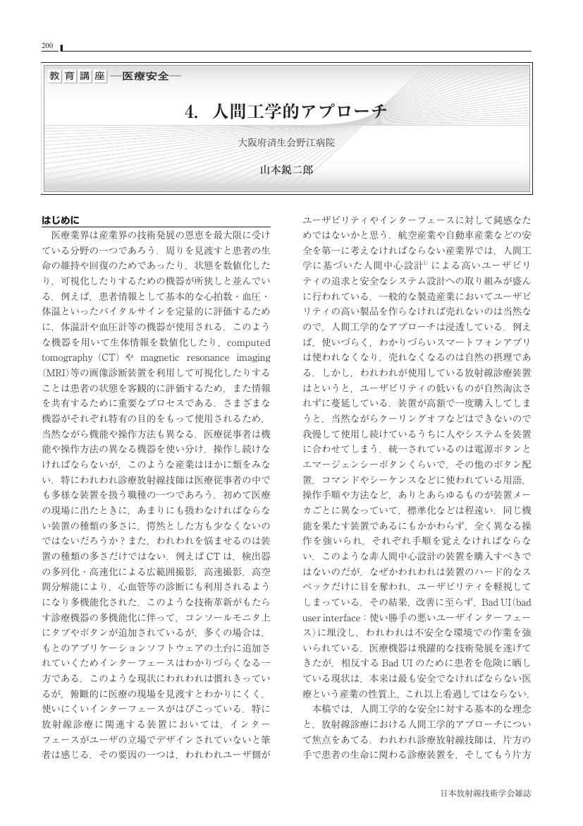
1 0 0 0 4. 人間工学的アプローチ
- 著者
- 山本 鋭二郎
- 出版者
- 公益社団法人 日本放射線技術学会
- 雑誌
- 日本放射線技術学会雑誌 (ISSN:03694305)
- 巻号頁・発行日
- vol.77, no.2, pp.200-211, 2021 (Released:2021-02-20)
- 参考文献数
- 13
- 著者
- 荒井 博史 久保 直樹 表 英彦 高橋 典子 勝浦 秀則 鈴木 幸太郎 古館 正従
- 出版者
- 公益社団法人 日本放射線技術学会
- 雑誌
- 日本放射線技術学会雑誌 (ISSN:03694305)
- 巻号頁・発行日
- vol.44, no.7, 1988
1 0 0 0 2 多目的カセッテホルダについて
- 著者
- 宮原 清英 岩田 康平
- 出版者
- 公益社団法人 日本放射線技術学会
- 雑誌
- 日本放射線技術学会雑誌 (ISSN:03694305)
- 巻号頁・発行日
- vol.54, no.9, 1998
1 0 0 0 エリアモニタ監視方式による感染症医療廃棄物の管理
- 著者
- 柳沢 正道 丸 繁勘 椎葉 眞一
- 出版者
- 公益社団法人 日本放射線技術学会
- 雑誌
- 日本放射線技術学会雑誌 (ISSN:03694305)
- 巻号頁・発行日
- vol.58, no.1, pp.130-132, 2002
- 参考文献数
- 4
- 被引用文献数
- 1 1
- 著者
- 笹本 和夫 長山 忠雄
- 出版者
- 公益社団法人 日本放射線技術学会
- 雑誌
- 日本放射線技術学会雑誌 (ISSN:03694305)
- 巻号頁・発行日
- vol.44, no.8, pp.1034, 1988-08-01 (Released:2017-06-28)
1 0 0 0 2.MRI の Radiomics
- 著者
- 酒井 晃二
- 出版者
- 公益社団法人 日本放射線技術学会
- 雑誌
- 日本放射線技術学会雑誌 (ISSN:03694305)
- 巻号頁・発行日
- vol.77, no.8, pp.866-875, 2021 (Released:2021-08-20)
- 参考文献数
- 103
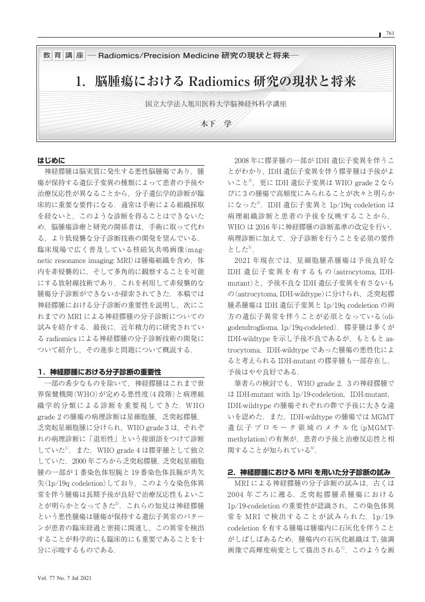
1 0 0 0 1.脳腫瘍における Radiomics 研究の現状と将来
- 著者
- 木下 学
- 出版者
- 公益社団法人 日本放射線技術学会
- 雑誌
- 日本放射線技術学会雑誌 (ISSN:03694305)
- 巻号頁・発行日
- vol.77, no.7, pp.761-764, 2021 (Released:2021-07-20)
- 参考文献数
- 16
- 著者
- 村松 千左子
- 出版者
- 公益社団法人 日本放射線技術学会
- 雑誌
- 日本放射線技術学会雑誌 (ISSN:03694305)
- 巻号頁・発行日
- vol.77, no.7, pp.760, 2021 (Released:2021-07-20)
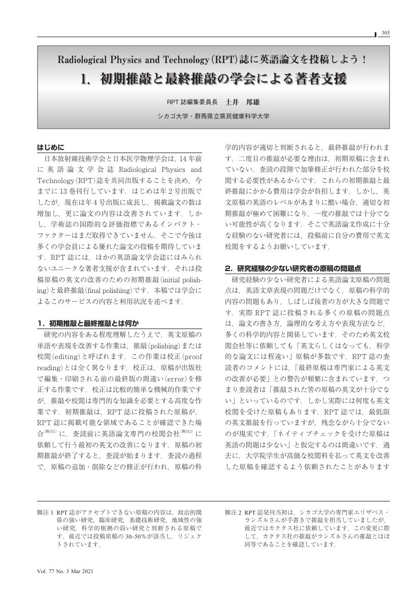
1 0 0 0 1. 初期推敲と最終推敲の学会による著者支援
- 著者
- 土井 邦雄
- 出版者
- 公益社団法人 日本放射線技術学会
- 雑誌
- 日本放射線技術学会雑誌 (ISSN:03694305)
- 巻号頁・発行日
- vol.77, no.3, pp.305-308, 2021 (Released:2021-03-20)
- 参考文献数
- 9
- 著者
- 菅原 時美
- 出版者
- 公益社団法人 日本放射線技術学会
- 雑誌
- 日本放射線技術学会雑誌 (ISSN:03694305)
- 巻号頁・発行日
- vol.21, no.4, 1966
- 著者
- 山城 晶弘 神谷 直紀 大塚 薫 駒津 和浩 伊東 洋一 久保田 展聡 小林 正人
- 出版者
- 公益社団法人 日本放射線技術学会
- 雑誌
- 日本放射線技術学会雑誌 (ISSN:03694305)
- 巻号頁・発行日
- vol.69, no.2, pp.163-169, 2013-02-20 (Released:2013-03-01)
- 参考文献数
- 13
- 被引用文献数
- 2
In magnetic resonance imaging (MRI), the ideal phantom should have similar T1 and T2 values to those of organs of interest for measuring the change in signal intensity, contrast ratio and contrast noise ratio. There have been several reports to develop such a phantom using materials with limited availability or complex methods. In this study, we have developed a simple phantom using indigestible dextrin and soluble calcium at 1.5-tesla MRI. The T1 and T2 values have been reduced by dissolving indigestible dextrin and soluble calcium in distilled water. The similar T1 and T2 values to those of organs (i.e., kidney cortex, kidney medulla, liver, spleen, pancreas, bone marrow, uterus myometrium, uterus endometrium, uterus cervix, prostate, brain white matter, and brain gray matter) have been obtained by varying the concentration of indigestible dextrin and soluble calcium. This phantom is easy to develop and has a potential to increase the accuracy of MRI phantom experiments.
1 0 0 0 OA 肝悪性腫瘍切除術前3DCTにおける門脈相撮影タイミングの最適化
- 著者
- 原田 耕平 千葉 彩佳 溝延 数房 沼澤 香夏子 今井 達也
- 出版者
- 公益社団法人 日本放射線技術学会
- 雑誌
- 日本放射線技術学会雑誌 (ISSN:03694305)
- 巻号頁・発行日
- vol.72, no.11, pp.1098-1104, 2016 (Released:2016-11-20)
- 参考文献数
- 12
- 被引用文献数
- 1
Preoperative three-dimensional computed tomography (3DCT) of the liver is the most important examination in performing preoperative simulation. Detailed visualization of the portal vein using the workstation is critical to enable accurate liver segmentation. However, the timing of imaging in the portal venous phase has mostly been reported equivalent to that of the liver screening examinations commonly performed. The purpose of this study was to examine the optimal timing of image capture to create the best portal vein visualization in preoperative 3DCT of the liver. Seventy-nine patients who underwent hepatectomy for malignant liver tumors were enrolled in this study. All patients were preoperatively examined using protocol A (imaging method separated into a portal venous phase and a hepatic venous phase) and then examined 1 week after surgery using protocol B (normal liver screening protocol). We first established the regions of interest in the portal vein and the hepatic vein and then compared CT values for these regions under protocol A and protocol B. The average CT value of the portal vein in protocol A and B was 239.8±28.1 HU and 202.2±18.5 HU, respectively. The average CT value of the portal vein in protocol A was significantly higher compared with protocol B (p<0.01). By introducing separate timing for portal venous phase imaging before preoperative 3DCT (protocol A), it is possible to satisfactorily depict the portal vein.
1 0 0 0 44. HALF BEAMを用いた腹部単純立位撮影 第二報
- 著者
- 田淵 真弘 松尾 孝人 小橋 義範 水内 敬枝
- 出版者
- 公益社団法人 日本放射線技術学会
- 雑誌
- 日本放射線技術学会雑誌 (ISSN:03694305)
- 巻号頁・発行日
- vol.51, no.3, 1995


