- 著者
- 田淵 真弘 松尾 孝人 小橋 義範 水内 敬枝
- 出版者
- 公益社団法人 日本放射線技術学会
- 雑誌
- 日本放射線技術学会雑誌 (ISSN:03694305)
- 巻号頁・発行日
- vol.50, no.10, 1994
1 0 0 0 OA インパルス応答を利用した核磁気共鳴画像のスライス厚測定
- 著者
- 山口 功 石森 佳幸 藤原 康博 谷内田 拓也 吉岡 千絵
- 出版者
- 公益社団法人 日本放射線技術学会
- 雑誌
- 日本放射線技術学会雑誌 (ISSN:03694305)
- 巻号頁・発行日
- vol.68, no.10, pp.1295-1306, 2012-10-20 (Released:2012-10-22)
- 参考文献数
- 13
- 被引用文献数
- 1 1
The purpose of this study was to verify the applicability of measurement of slice thickness of magnetic resonance imaging (MRI) by the delta method, and to discuss the measurement precision by the disk diameter and baseline setup of the slice profile of the delta method. The delta method used the phantom which put in the disk made of acrylic plastic. The delta method measured the full width at half maximum (FWHM) and the full width at tenth maximum (FWTM) from the slice profile of the disk signal. Evaluation of the measurement precision of the delta method by the disk diameter and baseline setup were verified by comparison of the FWHM and FWTM. In addition, evaluation of the applicability of the delta method was verified by comparison of the FWHM and FWTM using the wedge method. The baseline setup had the proper signal intensity of an average of 10 slices in the disk images. There were statistically significant difference in the FWHM between disk diameter of 10 mm and disk diameter of 30 mm and 5 mm. The FWHM of the disk diameter of 10 mm was smaller than the disk diameter of 30 mm and 5 mm. There was no statistically significant difference in the FWHM between the delta method and the wedge method. There is no difference in the effective slice thickness of the delta method and the wedge method. The delta method has an advantage in measurement of thin slice thickness.
1 0 0 0 OA 傾斜板法を用いた3D撮像のスライスプロファイル計測に対する一考察
- 著者
- 吉田 礼 町田 好男 小倉 隆英 田村 元 引地 健生 森 一生
- 出版者
- 公益社団法人 日本放射線技術学会
- 雑誌
- 日本放射線技術学会雑誌 (ISSN:03694305)
- 巻号頁・発行日
- vol.68, no.11, pp.1456-1466, 2012-11-20 (Released:2012-11-21)
- 参考文献数
- 16
- 被引用文献数
- 3 4
Recent progress in variable-flip-angle fast spin-echo technology has further extended the utility of three-dimensional (3D) magnetic resonance imaging (MRI) for clinical application. The slice profile in 3D MRI is the point spread function that has a sync form in principle, whereas a slice profile in 2D imaging provides information on characteristics of selective radio frequency excitation. We investigated the optimal condition to measure 3D slice profiles using a crossed thin-ramps phantom. We found that the profile data should cover a large area in order to evaluate both the main lobe and side lobes in the slice profile, and that the appropriate slice thickness was 2 mm. We also found that artifacts in the direction perpendicular to the slice create an offset error in the measured slice profile when 3D imaging. In this paper, we describe the optimal condition and some remarks on the slice profile evaluation for 3D MRI.
1 0 0 0 OA 放射線情報管理システムを用いた造影剤投与管理システムの有用性
- 著者
- 鈴木 伸忠 伊藤 肇 坂井 上之 越智 茂博 梁川 範幸
- 出版者
- 公益社団法人 日本放射線技術学会
- 雑誌
- 日本放射線技術学会雑誌 (ISSN:03694305)
- 巻号頁・発行日
- vol.76, no.5, pp.474-482, 2020 (Released:2020-05-20)
- 参考文献数
- 20
- 被引用文献数
- 1
We report on the construction of a system for managing prior information and injection condition used for contrast enhance CT examination using radiology information system (RIS). Contrast dose administration system using the RIS was possible to retrospectively investigate optimal injection conditions from the database. As the prior information, we designed the patientʼs profile information of the hospital information system (HIS) to reflect the patientʼs height, weight, and kidney function (eGFR, Cre), which is necessary information for contrast enhance CT examination, in the RIS. By adding E-Box (DICOM Gateway) to the injector, it became possible to reflect the amount of contrast agent used in patients and injection conditions at contrast enhance CT examination. The contrast agent use information is transmitted to RIS by using modality performed procedure step (MPPS). Database of injection condition at contrast enhance CT examination using the RIS, to determine the optimal injection conditions retrospectively. By utilizing the massive amount of clinical information stored in the RIS, the amount of contrast agent and injection condition at contrast enhance CT examination could be optimized. Reproducibility of the contrast effect can be secured. In the CE, evidence system linked with RIS, when considering the reproducibility at follow-up observation and comparative diagnosis in clinical practice, the contrast effect could be made constant. Contrast dose administration system using the RIS was useful.
1 0 0 0 OA 頭部 MRI 領域における深層学習のためのモーションアーチファクトジェネレータの開発
- 著者
- 塚本 ひかり 室 伊三男
- 出版者
- 公益社団法人 日本放射線技術学会
- 雑誌
- 日本放射線技術学会雑誌 (ISSN:03694305)
- 巻号頁・発行日
- vol.77, no.5, pp.463-470, 2021 (Released:2021-05-20)
- 参考文献数
- 8
- 被引用文献数
- 2
Purpose: We focused on deep learning for a reduction of motion artifacts in MRI. It is difficult to collect a large number of images with and without motion artifacts from clinical images. The purpose of this study was to create motion artifact images in MRI by simulation. Methods: We created motion artifact images by computer simulation. First, 20 different types of vertical pixel-shifted images were created with different shifts, and the amount of pixel shift was set from –10 to 10 pixels. The same method was used to create pixel-shifted images for horizontal shift, diagonal shift, and rotational shift, and a total of 80 types of pixel-shifted images were prepared. These images were Fourier transformed to create 80 types of k-space data. Then, phase encodings in these k-space data were randomly sampled and Fourier transformed to create artifact images. The reproducibility of the simulation images was verified using the deep learning network model of U-net. In this study, the evaluation indices used were the structural similarity index measure (SSIM) and peak signal-to-noise ratio (PSNR). Results: The average SSIM and PSNR for the simulation images were 0.95 and 31.5, respectively; those for the clinical images were 0.96 and 31.1, respectively. Conclusion: Our simulation method enables us to create a large number of artifact images in a short time, equivalent to clinical artifact images.
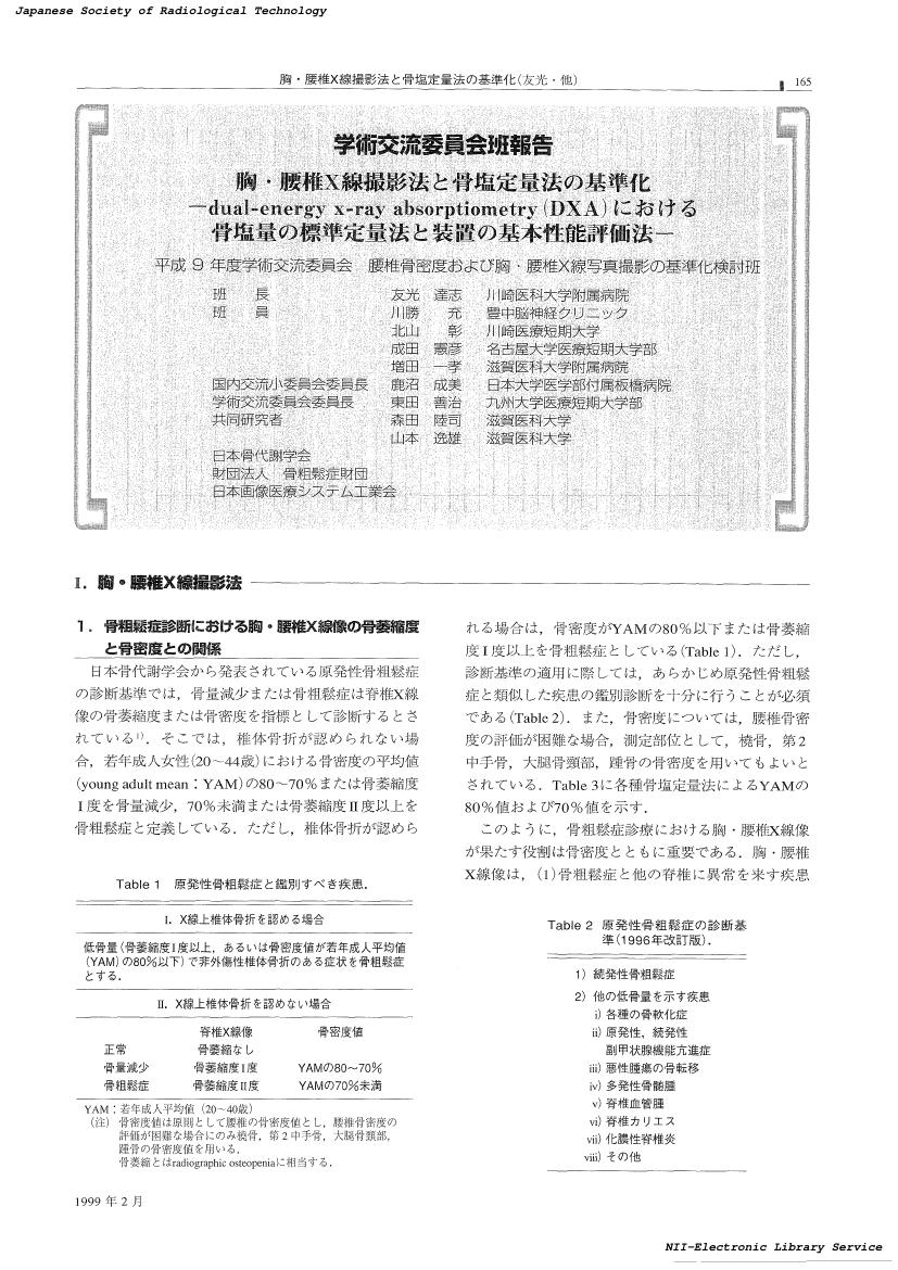
- 著者
- 友光 達志 川勝 充 北山 彰 成田 憲彦 増田 一孝 鹿沼 成美 東田 善治 森田 陸司 山本 逸雄
- 出版者
- 公益社団法人 日本放射線技術学会
- 雑誌
- 日本放射線技術学会雑誌 (ISSN:03694305)
- 巻号頁・発行日
- vol.55, no.2, pp.165-187, 1999-02-20 (Released:2017-06-30)
- 参考文献数
- 15
- 被引用文献数
- 1 1
1 0 0 0 OA 有鉤骨鉤基部撮影における撮影体位の検討
- 著者
- 髙松 俊介 宮川 誠一郎 佐藤 久弥 鈴木 航 西澤 剛 中村 雅美 梅田 宏孝 崔 昌五 加藤 京一 中澤 靖夫 池田 純
- 出版者
- 公益社団法人 日本放射線技術学会
- 雑誌
- 日本放射線技術学会雑誌 (ISSN:03694305)
- 巻号頁・発行日
- vol.70, no.6, pp.549-555, 2014 (Released:2014-06-20)
- 参考文献数
- 13
The hamate bone, one of the carpal (wrist) bones, has a large uncinate process protruding from the palm side. In sports such as golf and tennis, the hamate bone can break if is subjected to a high external force, such as from the handle of a racquet or club. At our hospital we take X-ray images of the hamate bone from two directions: an axial image through the carpal tunnel and an image at the base of the hamate hook (conventional method). While the conventional method makes it easy to create images of the base of the hamate hook, the patient may suffer pain during image-taking because the hamate bone is pulled to cause radial flexion. We therefore investigated a method of imaging that would create three-dimensional computed tomography (3DCT) images of the base of the hamate hook in which the patient would only have to only rotate the wrist externally and elevate the fore-arm without any radial flexion. Our results suggest that it is possible to obtain images of the base of the hamate hook as clear as those acquired using the conventional method with the patient in a comfortable and painless position taking images at an external rotation angle of 50.3° and a forearm elevation angle of 20.3°.
- 著者
- 山田 澄人 山下 清司
- 出版者
- 公益社団法人 日本放射線技術学会
- 雑誌
- 日本放射線技術学会雑誌 (ISSN:03694305)
- 巻号頁・発行日
- vol.50, no.8, 1994
1 0 0 0 OA 胸部 CT 撮影における wide volume scan 使用時の被ばく特性評価
- 著者
- 酒向 健二 松原 孝祐 松本 真
- 出版者
- 公益社団法人 日本放射線技術学会
- 雑誌
- 日本放射線技術学会雑誌 (ISSN:03694305)
- 巻号頁・発行日
- vol.77, no.3, pp.284-292, 2021 (Released:2021-03-20)
- 参考文献数
- 22
Purpose: A volume scan can cover a range of 160 mm with a single gantry rotation. It can be performed sequentially (a wide volume [WV] scan) to cover more than 160 mm, and volume Xact+ (Xact+) can be used when volume scan is done to extend the reconstruction area. The purpose of this study was to investigate the dose distribution and organ doses for a WV scan during chest CT. Method: We arranged radiophotoluminescence glass dosimeters (RPLDs) linearly on the surface and inside of the phantom to evaluate the dose distribution along the z-axis. We also placed RPLDs at the lens, thyroid, and breast positions to evaluate organ doses. We performed WV and helical scans and WV scan using Xact+. Result: The absorbed doses increased at the borders of the volume scans, and dose peaks were observed there. The organ doses for the WV scan outside the acquisition range were lower than those for the helical scan. The organ doses inside the acquisition range changed by the locations of borders. Conclusion: The WV scan increases the absorbed doses at the overlapping scanned regions, which can be reduced by using Xact+.
- 著者
- 尾崎 秀道 影山 勉 村木 威
- 出版者
- 公益社団法人 日本放射線技術学会
- 雑誌
- 日本放射線技術学会雑誌 (ISSN:03694305)
- 巻号頁・発行日
- vol.41, no.5, 1985
1 0 0 0 OA CT-AEC を用いた低管電圧撮影の被ばくに関する検討
- 著者
- 髙田 光雄 松原 孝祐 越田 吉郎 太郎田 融
- 出版者
- 公益社団法人 日本放射線技術学会
- 雑誌
- 日本放射線技術学会雑誌 (ISSN:03694305)
- 巻号頁・発行日
- vol.71, no.4, pp.332-337, 2015 (Released:2015-04-20)
- 参考文献数
- 14
- 被引用文献数
- 2 5
The purpose of our study was to investigate radiation dose for lower tube voltage CT using automatic exposure control (AEC). An acrylic body phantom was used, and volume CT dose indices (CTDIvol) for tube voltages of 80, 100, 120, and 135 kV were investigated with combination of AEC. Average absorbed dose in the abdomen for 100 and 120 kV were also measured using thermoluminescence dosimeters. In addition, we examined noise characteristics under the same absorbed doses. As a result, the exposure dose was not decreased even when the tube voltage was lowered, and the organ absorbed dose value became approximately 30% high. And the noise was increased under the radiographic condition to be an equal absorbed dose. Therefore, radiation dose increases when AEC is used for lower tube voltage CT under the same standard deviation (SD) setting with 120 kV, and the optimization of SD setting is crucial.
1 0 0 0 OA 脳動脈瘤クリッピング術後の血管描出を目的とした超短TE シーケンスの有用性
- 著者
- 田久保 早紀 川﨑 康平 長渡 努 松本 正信 景山 貴洋
- 出版者
- 公益社団法人 日本放射線技術学会
- 雑誌
- 日本放射線技術学会雑誌 (ISSN:03694305)
- 巻号頁・発行日
- vol.76, no.2, pp.177-184, 2020 (Released:2020-02-20)
- 参考文献数
- 15
- 被引用文献数
- 1 7
The aims of this study were to elucidate signal pattern of cerebral aneurysm clip in brain magnetic resonance angiography (MRA) using non-contrast enhanced ultra-short echo time (UTE) sequence and to explore effective utilization of this novel technique for patients, who underwent cerebral aneurysm clipping. The clip was embedded in homemade phantom and scanned using UTE sequence. We investigated characteristic features of the artifacts derived from the clip. Besides, we compared the volume of signal loss between conventional time-of-flight (TOF) and UTE-MRA in 50 patients with the cerebral aneurysm clip. In phantom study, the clip was delineated as signal void area fully surrounded by high signal on original images. On reconstructed short-axial views for the clip, four-leaf clover pattern of artifact was observed when clip was arranged orthogonal to the static magnetic field. On the other hand, this artifact disappeared when the clip was arranged in parallel with the static magnetic field. The volume of signal loss in clinical cases was significantly reduced in UTE-MRA (P < 0.05): 1.30, 0.52–2.77 cm3 for TOF; 0.84, 0.28–1.74 cm3 for UTE (median, range). The scan time for UTE-MRA was 2 minutes and 52 seconds. To understand the characteristic feature of the artifacts from the clip could contribute to define vascular structure in image interpretation. Adding UTE-MRA to routine protocol is useful approach for follow-up imaging after cerebral aneurysm clipping with clinically acceptable prolongation of the scan time.
1 0 0 0 (9) メディカルボイス (音声合成発生器) の改良
- 著者
- 本間 龍夫 三浦 正和 藤井 博美
- 出版者
- 公益社団法人 日本放射線技術学会
- 雑誌
- 日本放射線技術学会雑誌 (ISSN:03694305)
- 巻号頁・発行日
- vol.48, no.2, 1992
日本においても海外交流が栄んになり、商品輸出入のみでなく、旅行者、研修生という外国人の入国がふえました。特に近隣の中国人、韓国人や南米2世のブラジル人等の人々が病院で治療をうけたり、検診をうける機会がふえましたが、各国語を話せる病院スタッフは大変少ないと思われます。その折X線室で撮影する先生方の替わりに外国語で話す音声装置があると便利である。又、耳の不自由な人や老人のペイシエントケア用品として表示器もあるが、CTガントリー内で寝ていても見えるように改良する必要があるという施設があった。
1 0 0 0 OA 金属アーチファクト低減処理画像に対する逐次近似応用再構成の影響
- 著者
- 保吉 和貴 佐藤 俊光 岡田 明男
- 出版者
- 公益社団法人 日本放射線技術学会
- 雑誌
- 日本放射線技術学会雑誌 (ISSN:03694305)
- 巻号頁・発行日
- vol.74, no.8, pp.797-804, 2018 (Released:2018-08-20)
- 参考文献数
- 22
- 被引用文献数
- 1 1
Purpose: This study aimed to evaluate the effect of adaptive iterative dose reduction 3D (AIDR 3D) on the computed tomography (CT) image quality by using single energy metal artifact reduction (SEMAR). Materials & Methods: A water phantom (22 cmφ) with the stem for total hip arthroplasty made of titanium was scanned. The volume CT dose index (CTDIvol) was set to 8.9 and 5.0 mGy. The reconstruction was performed using filtered back projection and AIDR 3D by soft kernel (FC13) and SEMAR. The averaged profile method was used for the quantitative evaluation of artifacts. We placed a rectangular region-of-interest on the artifact part, and obtained the x-direction averaged profile (Profile A). Profile B was obtained using a water phantom without metal. Profiles A and B were normalized as Profiles A′ and B′ using the mean value calculated from Profile B. Based on the standard deviation (SD) calculated from Profile B′, the background variation level was defined as ±2SD, and subtracted from Profile A′ (Profile A″). Finally, the area of Profile A″ was calculated and defined as Artifacttotal. Artifactover, and Artifactunder, respectively, the positive- and negative-side components of Artifacttotal. Results: Both Artifacttotal and Artifactunder increased according to the strength of AIDR 3D. The variations of Artifactover and Artifactunder, due to the AIDR 3D strength, were small and large, respectively. Further, in comparison with a high dose, the effect of artifact emphasis increased at low dose. Therefore, it should be noted that stronger AIDR 3D can emphasize the residual metal artifact.
1 0 0 0 31 銀塩熱現像方式ドライレーザーイメージャ2機種の比較について
- 著者
- 高橋 淳 小川 清 松田 恵雄 庭田 清隆
- 出版者
- 公益社団法人 日本放射線技術学会
- 雑誌
- 日本放射線技術学会雑誌 (ISSN:03694305)
- 巻号頁・発行日
- vol.56, no.9, 2000
- 著者
- 加藤 清
- 出版者
- 公益社団法人 日本放射線技術学会
- 雑誌
- 日本放射線技術学会雑誌 (ISSN:03694305)
- 巻号頁・発行日
- vol.10, 1955
- 著者
- 佐川 良 後藤 正美 秋山 晋
- 出版者
- 公益社団法人 日本放射線技術学会
- 雑誌
- 日本放射線技術学会雑誌 (ISSN:03694305)
- 巻号頁・発行日
- vol.43, no.11, 1987
- 著者
- 石屋 博樹 千田 浩一 斎 政博 佐藤 弘之 有馬 宏寧 佐々木 正寿
- 出版者
- 公益社団法人 日本放射線技術学会
- 雑誌
- 日本放射線技術学会雑誌 (ISSN:03694305)
- 巻号頁・発行日
- vol.49, no.8, 1993
透視条件下において、X線アナライザの管電圧測定値と実測値はほぼ一致した。また、線量率測定に於いて、X線アナライザ2機種共に低い管電圧側で基準線量計の値より低い値を示し、管電圧が高くなる程、基準線量計の値に近づいた。今回の測定では、低い管電圧の時、線量率及び管電圧の測定が不能になったり、線量率の測定値が低くなったりするものもあったが、この原因としてはX線アナライザの測定レンジの下限を越えたためか、又は、検出限界ぎりぎりのところで測定したために起こったものと考えられる。以上より、X線アナライザは、透視条件という低いX線出力下においても、測定レンジの範囲内では充分QCに使用可能であると思われた。
1 0 0 0 OA DR システムにおけるDQE 測定時の各因子の測定精度に関する検討
- 著者
- 國友 博史 小山 修司 東出 了 市川 勝弘 服部 真澄 岡田 陽子 林 則夫 澤田 道人
- 出版者
- 公益社団法人 日本放射線技術学会
- 雑誌
- 日本放射線技術学会雑誌 (ISSN:03694305)
- 巻号頁・発行日
- vol.70, no.7, pp.653-661, 2014 (Released:2014-07-23)
- 参考文献数
- 23
In the detective quantum efficiency (DQE) evaluation of detectors for digital radiography (DR) systems, physical image quality indices such as modulation transfer function (MTF) and normalized noise power spectrum (NNPS) need to be accurately measured to obtain highly accurate DQE evaluations. However, there is a risk of errors in these measurements. In this study, we focused on error factors that should be considered in measurements using clinical DR systems. We compared the incident photon numbers indicated in IEC 62220-1 with those estimated using a Monte Carlo simulation based on X-ray energy spectra measured employing four DR systems. For NNPS, influences of X-ray intensity non-uniformity, tube voltage and aluminum purity were investigated. The effects of geometric magnifications on MTF accuracy were also examined using a tungsten edge plate at distances of 50, 100 and 150 mm from the detector surface at a source-image receptor distance of 2000 mm. The photon numbers in IEC 62220-1 coincided with our estimates of values, with error rates below 2.5%. Tube voltage errors of approximately ±5 kV caused NNPS errors of within 1.0%. The X-ray intensity non-uniformity caused NNPS errors of up to 2.0% at the anode side. Aluminum purity did not affect the measurement accuracy. The maximum MTF reductions caused by geometric magnifications were 3.67% for 1.0-mm X-ray focus and 1.83% for 0.6-mm X-ray focus.
1 0 0 0 汝,英語を恐れることなかれ
- 著者
- 白石 順二
- 出版者
- 公益社団法人 日本放射線技術学会
- 雑誌
- 日本放射線技術学会雑誌 (ISSN:03694305)
- 巻号頁・発行日
- vol.72, no.5, pp.I, 2016
