- 著者
- 冨永 和宏 松尾 長光 上野 功 森本 守 重本 康晴 空閑 祥浩 水野 明夫 佐々木 元賢
- 出版者
- 社団法人 日本口腔外科学会
- 雑誌
- 日本口腔外科学会雑誌 (ISSN:00215163)
- 巻号頁・発行日
- vol.39, no.8, pp.879-890, 1993
- 被引用文献数
- 1 1
To evaluate the utility of subperiosteal tissue expansion (STE) for mandibular augmentation with hydroxylapatite (HA) particles, two experimental studies in adult mongrel dogs were performed. One was to observe histologic changes following STE and to compare the tissue response to STE with that to subcutaneous expansion. The other was to observe clinical and histologic changes following implantation of HA particles in the subperiosteal expanded bed. In the first experiment, small tissue expanders were placed and inflated subperiosteally in the mandible and subcutaneously in the breast. A fibrous connective tissue capsule was formed surrounding the implanted expanders. Periosteum was replaced by simple connective tissue during expansion. Capsule formation during STE was much more rapid than the subcutaneous expansion. In the subperiosteal group, a thick fibrous capsule consisting of coarse collagen fibers was observed one week after full inflation. Leaving the fully inflated subperiosteal expanders in place more than one month accelerated the resorption of the underlying bone. In the second experiment, one week after full expansion of the mandibular subperiosteal expander, the expander was removed and HA particles were implanted in the expanded bed. Despite marked augmentation with excessive amounts of HA particles, there was no deformity or infection of the grafts, which maintained their designed controur. The firm fibrous capsule formed by STE permitted consolida tion of HA particles easily and effectively, and prevented migration of the particles. Immobilization of the grafts was achieved within two months following implantation of the HA particles.
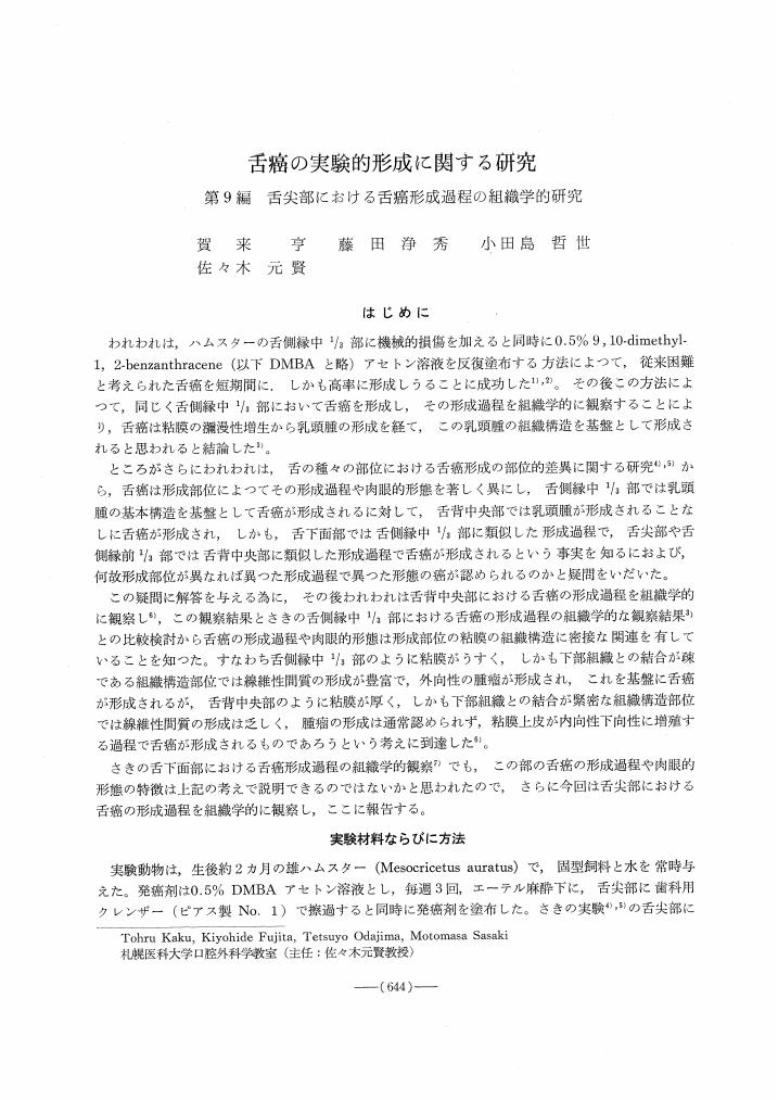
1 0 0 0 OA 舌癌の実験的形成に関する研究
- 著者
- 賀来 亨 藤田 浄秀 小田島 哲世 佐々木 元賢
- 出版者
- 特定非営利活動法人 日本口腔科学会
- 雑誌
- 日本口腔科学会雑誌 (ISSN:00290297)
- 巻号頁・発行日
- vol.23, no.4, pp.644-654, 1974 (Released:2011-09-07)
- 参考文献数
- 7
1 0 0 0 血友病B患者における抜歯例
- 著者
- 千野 武広 田中 俊彦 針谷 竜宜 藤井 久弥 佐々木 元賢 羽田 靖子 黒川 一郎
- 出版者
- Japanese Stomatological Society
- 雑誌
- 日本口腔科学会雑誌 (ISSN:00290297)
- 巻号頁・発行日
- vol.20, no.3, pp.559-566, 1971
