1 0 0 0 OA 院内製剤中のエンドトキシン測定と荷電フィルタの性能評価
- 著者
- 大石 雅子 勝浦 正人 片岡 和三郎 黒川 信夫
- 出版者
- 一般社団法人日本医療薬学会
- 雑誌
- 医療薬学 (ISSN:1346342X)
- 巻号頁・発行日
- vol.27, no.5, pp.452-460, 2001-10-10 (Released:2011-03-04)
- 参考文献数
- 12
- 被引用文献数
- 1 1
The prevention of endotoxin (ETX) contamination is important for manufacturing injections. As a result, the concentration of ETX in injections has been recently regulated in the Japanese Pharmacopoeia. In this study, ETX in 8 injections prepared at a hospital pharmacy were measured and the removal of ETX by filtration using a charged membrane filter (CMF) made of Nylon 66 (Zetapor®, CUNO) was evaluated. ETX was automatically measured with a Limulus test employing a turbidmetric kinetic assay (Toxinometer®, WAKO). ETX in H2SeO3 and MnCl2 injections were measured without dilution but others need to be diluted to eliminate any interference with the reaction. In particular, the ZnSO4 injection was diluted 2000 times. In the preparations, only 1 % indigocarmin injection showed 0.311 EU/mL of ETX.In a preliminary evaluation, CMF removed ETX completely in the non-electrolytic solutions such as glucose. However, in electrolytic solutions like NaCl, the filtration efficiency of CMF was suggested to decrease by some factors such as the concentration of electrolytes, the pH of the solution and the origin of ETX. In 0.9% NaCl solution to which the control standard ETX was added, the recovery rate of ETX using CMF was 0.3-2%. In 10% NaCl solution, the recovery rate was 37-47% under the same conditions, but it became 75-79% when another origin of ETX was added. In the actual process of preparations, ETX was found in 1 % indigocarmin injection, but it was not found in them when CMF was used. ETX was not removed by a usual filter membrane without any charging and not inactivated completely by steam sterilization. Therefore, filtration using CMF was found to effectively remove ETX together with steam sterilization.Filtration using CMF is thus considered to be a simple and effective method for maintaining a good quality of injections prepared at hospital pharmacies.
1 0 0 0 OA 薬剤部製剤室の空気清浄度による環境評価と浮遊粒子数に関する影響因子の解析
- 著者
- 大石 雅子 片岡 和三郎 中川 知子 勝浦 正人 池田 賢二 黒川 信夫
- 出版者
- 一般社団法人日本医療薬学会
- 雑誌
- 医療薬学 (ISSN:1346342X)
- 巻号頁・発行日
- vol.27, no.3, pp.212-220, 2001-06-10 (Released:2011-03-04)
- 参考文献数
- 9
In this study, the present state of the air cleanliness in the drug preparation room at a hospital pharmacy was evaluated, and factors affecting airborne particle numbers (APN) such as the number and the movement of workers and the materials on working clothes and cloths were investigated. In addition, the effect of environmental conditions on air cleanliness on a clean bench was compared. APN was measured with an Aerosol Particle Counter.The maximum 0.5μm APN values while working in the aseptic preparation room were 3, 610, 1, 312 (less than 10, 000 in GMP) and the non-aseptic room were 8, 008, 2, 660 (less than 100, 000) respectively. The conditions of all rooms were sufficiently suitable for drug preparation according to the criteria of GMP.Concerning factors affecting APN, the movement of workers increased the APN much more than the number of workers. The degree of dispersing particles differed greatly depending on the materials of the working clothes and cloths. A decrease to less than 1 /100 can be obtained by the selection of suitable materials for working clothes such as Overall made of polypropylene non-woven fabric from which few of fibers disperse. It is remarkable that smaller particles are dispersed from clothes even after passing through an air shower. In addition, it was confirmed that the dispersing of particles from cloths and rags was also a problem.As long as prescribed methods were used for the clean bench, the air cleanliness inside the clean bench was kept sufficient even through the external air conditions or locations were not so clean.
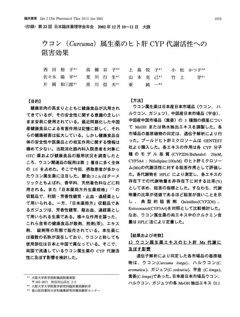
1 0 0 0 OA ウコン (Curcuma) 属生薬のヒト肝CYP代謝活性への阻害効果
1 0 0 0 OA 外来がん化学療法部門システムの追加導入と混合調製業務に寄与する因子の多変量解析
- 著者
- 池田 賢二 竹上 学 但馬 重俊 宮脇 康至 東 真樹子 八木 悠理子 亀川 秀樹 黒川 信夫
- 出版者
- 一般社団法人日本医療薬学会
- 雑誌
- 医療薬学 (ISSN:1346342X)
- 巻号頁・発行日
- vol.32, no.5, pp.436-444, 2006-05-10 (Released:2007-11-09)
- 参考文献数
- 15
- 被引用文献数
- 3 3
Pharmaceutical support systems for anti-neoplastic drug preparation for chemotherapy in hospital information systems are in need of improvement. With this in mind we developed a new support system in collaboration with system engineers (SEs) in January 2005. In the presentation of our requirements for the support system to the SEs, the most important part of the development process, we proposed a development model based on an end-user prototype system. The model comprised a five-step design and development of the prototype system through end-user computing, evaluation of the prototype system through actual use, the introduction of a step to determine the exact task requirements, detailed discussions with the SEs, and installation. Among the functions of the prototype system were support functions for the input of prescriptions, inventory management and task data assessment and a print-out function for record sheets. This model produced excellent results for the upgrade whose express purpose was the introduction of an end-user computing step. Furthermore, to evaluate the preparation support system, time-determinative factors involved in the preparation were analyzed and compared using data collected in November 2004 and March 2005. In the evaluation, two pharmacists dispensed a total of 791 and 1003 admixtures for 341 and 426 injection prescriptions, respectively, and the average preparation times were 18.15 and 15.43 minutes/prescription, respectively. Multiple regression analysis revealed several time-determinative factors with correlation coefficients of 0.803 and 0.668. The significant time-determinative factors were the number of vials, ampoules, and bottles for admixtures and the properties of drugs dispensed. The new system has resulted in improved patient safety and operational efficiency.
1 0 0 0 OA 薄層クロマトグラフィーを用いるアマチャ中のd-phyllodulcinのけい光定量法
- 著者
- 萩原 義郎 小西 逞夫 黒川 信夫 橋本 庸平 立花 陽子
- 出版者
- 日本生薬学会
- 雑誌
- 生薬学雑誌 (ISSN:00374377)
- 巻号頁・発行日
- vol.28, no.1, pp.47-50, 1974-06-20
A new method for the determination of d-phyllodulcin in Sweet Hydrangea by fluorometric measurements was established. After its seperation from other constituents by TLC and exposed to ultraviolet ray of 263.5 mμ, the d-phyllodulcin portion seperated by TLC gives blue fluorescence. By measurment of fluorescence intensity, d-phyllodulicin is determined quantitatively in concentration range of 1.0-4.0 μg/ml.

