1 0 0 0 OA 日本医科大学の名のもとに
- 著者
- 大塚 茂
- 出版者
- 日本医科大学医学会
- 雑誌
- 日本医科大学医学会雑誌 (ISSN:13498975)
- 巻号頁・発行日
- vol.10, no.2, pp.121-122, 2014 (Released:2014-05-08)
1 0 0 0 OA 基礎科学教育の今後
- 著者
- 野村 俊明
- 出版者
- 日本医科大学医学会
- 雑誌
- 日本医科大学医学会雑誌 (ISSN:13498975)
- 巻号頁・発行日
- vol.7, no.4, pp.166-168, 2011 (Released:2011-12-22)
- 参考文献数
- 14
1 0 0 0 OA 乳房増大術後遺症―シリコンと炭化水素系物質の重複注入例の画像診断
- 著者
- 百束 比古 水野 博司 汲田 伸一郎
- 出版者
- 日本医科大学医学会
- 雑誌
- 日本医科大学医学会雑誌 (ISSN:13498975)
- 巻号頁・発行日
- vol.6, no.2, pp.56-57, 2010 (Released:2010-05-06)
- 参考文献数
- 3
1 0 0 0 OA 花ざかりの森から
- 著者
- 小平 祐造
- 出版者
- 日本医科大学医学会
- 雑誌
- 日本医科大学医学会雑誌 (ISSN:13498975)
- 巻号頁・発行日
- vol.11, no.1, pp.41-42, 2015-02-15 (Released:2015-03-06)
- 著者
- 岩切 勝彦 田中 由理子
- 出版者
- 日本医科大学医学会
- 雑誌
- 日本医科大学医学会雑誌 (ISSN:13498975)
- 巻号頁・発行日
- vol.6, no.1, pp.4-6, 2010 (Released:2010-03-05)
- 被引用文献数
- 1
1 0 0 0 OA 認知症の鑑別診断
- 著者
- 山崎 峰雄
- 出版者
- 日本医科大学医学会
- 雑誌
- 日本医科大学医学会雑誌 (ISSN:13498975)
- 巻号頁・発行日
- vol.8, no.4, pp.274-279, 2012 (Released:2012-12-28)
- 参考文献数
- 4
1 0 0 0 OA 街ぐるみ認知症相談センターの4年間の活動状況
- 著者
- 石渡 明子 北村 伸 野村 俊明 根本 留美 石井 知香 若松 直樹 片山 泰朗 川並 汪一
- 出版者
- 日本医科大学医学会
- 雑誌
- 日本医科大学医学会雑誌 (ISSN:13498975)
- 巻号頁・発行日
- vol.9, no.1, pp.14-19, 2013 (Released:2013-03-11)
- 参考文献数
- 11
Aim: Community Consultation Center was established in 2007 as a core facility of a project entitled "Community Support Network for Citizens with Mild Cognitive Impairment and Dementia" subsidized by the Ministry of Education, Culture, Sports, Science and Technology. This study reports the activity within the facility and users' outcome. Methods: At the facility, users consulted their memory problem, and a screening tool with a touch-panel type computer (TP) was used to check their memory loss. Dementia was suspected when the TP score was 12 or less points, and clinical psychotherapist implemented Mini-Mental state examination. All the results were summarized in reports, and we prompted users to see their primary doctors, or nearby medical institutes that we offered. In this study, we asked these medical institutes of their outcome. Informed consent was obtained from all users. Results: A total of 2,802 people visited the Center, and 1,565 people registered (male/female=519/1,046; mean age, 74 years). 561 people used the center twice or more. Among 1,354 who had TP, 722 users got a score under 12 (46.1%). A total of 409 responses from medical institutes were collected. The data revealed that Mild cognitive impairment (MCI) was 11.2%, Alzheimer's disease was 37.1%, and vascular dementia was 8.00%. Conclusion: These results indicate that approximately half of the users of the Center was suspected dementia, and a prevalence of both MCI and dementia reached to about 60%. This Center has proven to be useful for early detection and diagnosis.
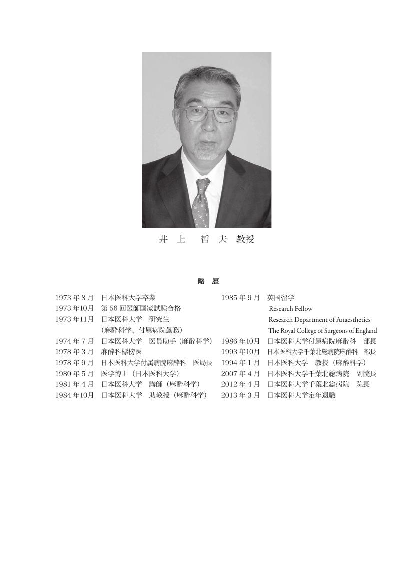
1 0 0 0 OA 気道へのアプローチ
- 著者
- 井上 哲夫
- 出版者
- 日本医科大学医学会
- 雑誌
- 日本医科大学医学会雑誌 (ISSN:13498975)
- 巻号頁・発行日
- vol.9, no.2, pp.69-76, 2013 (Released:2013-05-08)
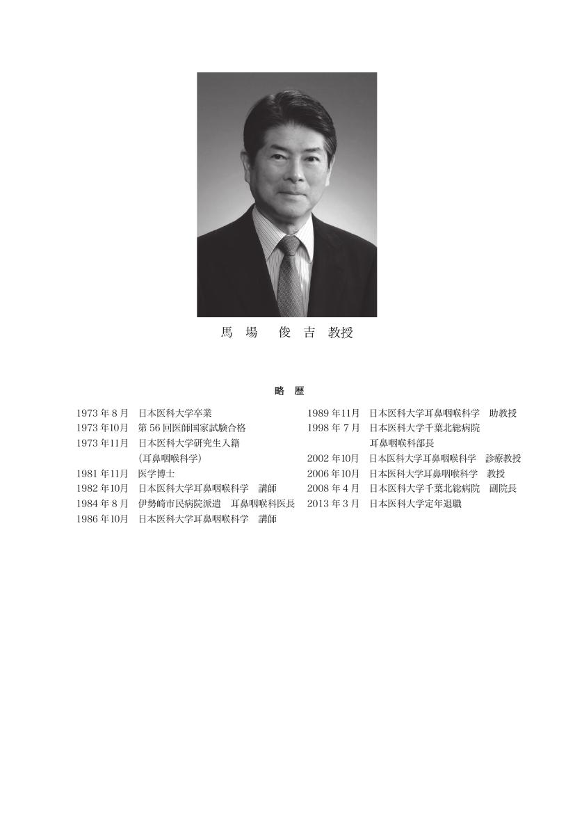
1 0 0 0 OA 耳鳴の治療について
- 著者
- 馬場 俊吉
- 出版者
- 日本医科大学医学会
- 雑誌
- 日本医科大学医学会雑誌 (ISSN:13498975)
- 巻号頁・発行日
- vol.9, no.2, pp.54-60, 2013 (Released:2013-05-08)
- 著者
- 澤 芳樹
- 出版者
- 日本医科大学医学会
- 雑誌
- 日本医科大学医学会雑誌 (ISSN:13498975)
- 巻号頁・発行日
- vol.5, no.1, pp.22-26, 2009 (Released:2009-04-15)
- 参考文献数
- 7
1 0 0 0 OA 2.組織細胞化学シリーズ(若手研究者へのヒント) 免疫電子顕微鏡法の基礎(3)
- 著者
- 小澤 一史 松崎 利行
- 出版者
- 日本医科大学医学会
- 雑誌
- 日本医科大学医学会雑誌 (ISSN:13498975)
- 巻号頁・発行日
- vol.5, no.4, pp.215-220, 2009 (Released:2009-11-17)
- 参考文献数
- 9
- 被引用文献数
- 1 1
Immunohistochemistry is concerned with the detection of specific biological substances at the light and electron microscopic levels with antibodies labeled with visible markers, such as horseradish peroxidase and colloidal gold. In particular, the immunohistochemistry of electron microscopy has provided much morphological and biological information. Immunoelectron microscopy can be classified into three methods, i.e., pre-embedding, postembedding, and nonembedding methods, on the basis of the step during which the immunoreaction is applied to the biological specimens. Each method has both advantages and disadvantages, so we should select the method according to the biological purpose. An overview of immunoelectron microscopy is given, and several electron micrographs using immunohistochemical techniques are shown.
1 0 0 0 OA 食道・胃静脈瘤治療
- 著者
- 吉田 寛 真々田 裕宏 谷合 信彦 山下 精彦 田尻 孝
- 出版者
- 日本医科大学医学会
- 雑誌
- 日本医科大学医学会雑誌 (ISSN:13498975)
- 巻号頁・発行日
- vol.1, no.4, pp.161-167, 2005 (Released:2005-11-09)
- 参考文献数
- 67
Bleeding from esophagogastric varices is a catastrophic complication of chronic liver disease. We have been attempted surgery, embolization, and endoscopic treatment for the treatment of esophagogastric varices. Endoscopic injection sclerotherapy (EIS) is an established treatment for esophageal varices. EIS is associated with a high incidence of local and systemic complications. Endoscopic variceal ligation (EVL) is increasingly used because of its safety and simplicity and because no sclerosant is used. Nevertheless, EVL is not always effective, and early recurrences have been reported. Furthermore, most patients with esophageal varices treated endoscopically require treatment for recurrent varices. We invented that EVL performed three times at bimonthly intervals. EVL performed at bimonthly intervals for the treatment of esophageal varices attained a higher complete eradication rate, a lower recurrence rate, and a lower additional treatment rate. It is generally believed that bleeding from gastric varices is more severe than bleeding from esophageal varices, but bleeding from gastric varices occurs less commonly than from esophageal varices. The endoscopic risk factors for bleeding from esophageal varices include presence of raised red markings, cherry-red spots, blue color, and large size. However the risk factors for bleeding from gastric varices have yet to be characterized. Once gastric variceal hemorrhage did occur, bleeding from these varices was successfully stopped in all cases. Therefore, prophylactic treatment of gastric varices is not recommended.
1 0 0 0 OA 2.臨床におけるFDG‐PET検査 脳疾患におけるFDG-PET検査(IV)
- 著者
- 水村 直 汲田 伸一郎
- 出版者
- 日本医科大学医学会
- 雑誌
- 日本医科大学医学会雑誌 (ISSN:13498975)
- 巻号頁・発行日
- vol.3, no.3, pp.125-127, 2007 (Released:2007-07-12)
- 被引用文献数
- 1


