5 0 0 0 OA 難治性虹彩炎を伴ったTattoo sarcoidosis(刺青サルコイドーシス)の1例
- 著者
- 吉田 秀 遠藤 平仁 西 正大 飯塚 進子 木村 美保 田中 住明 石川 章 近藤 啓文
- 出版者
- 一般社団法人 日本臨床リウマチ学会
- 雑誌
- 臨床リウマチ (ISSN:09148760)
- 巻号頁・発行日
- vol.18, no.3, pp.225-231, 2006-09-30 (Released:2016-12-30)
- 参考文献数
- 16
Tattoo sarcoidosis is a rare clinical entity that shows a sarcoidosis like granuloma developing from tattoo skin and clinical symptoms of systemic sarcoidosis after long term courses of tattoos. Hypersensitivity reaction for metal included in a pigment of tattoo pigment may assume to be the cause of tattoo sarcoidosis. A man aged 20s developed symptoms similar to those of systemic sarcoidosis after receiving a multicolored tattoo covering the skin of his entire body. He put a tattoo (red, green, black, gray, yellow and gray) over his entire body 10 years ago. There were severely painful subcutaneous masses about 1 cm in size on a brown and green tattoo on back and lower extremities. A biopsy of the part of the nodule showed a foreign body granuloma containing multinuclear giant cells. Photophobia and decreased visual acuity developed afterwards, and he was diagnosed with bilateral iritis. In addition, hilar lymphadenopathy was founded in a chest X-ray and CT, persistent fever also showed. We diagnosed it as sarcoid reaction by the tattoo because of skin epithelioid granuloma, iritis, and bilateral hilar lympadenopathy in CT. After hospitalization, his eyes were injected with dexamethazone and he took oral prednisolone 40 mg/day for iritis. Severe iritis was not improved by administration of only steroids and improved in combinating azathioprine. We report this as the interesting case that showed a sarcoidosis like reaction to the pigment of a tattoo.
2 0 0 0 膠原病に伴う心外膜炎・心囊液貯留
- 著者
- 遠藤 平仁
- 出版者
- 医学書院
- 雑誌
- 呼吸と循環 (ISSN:04523458)
- 巻号頁・発行日
- vol.63, no.11, pp.1037-1041, 2015-11-15
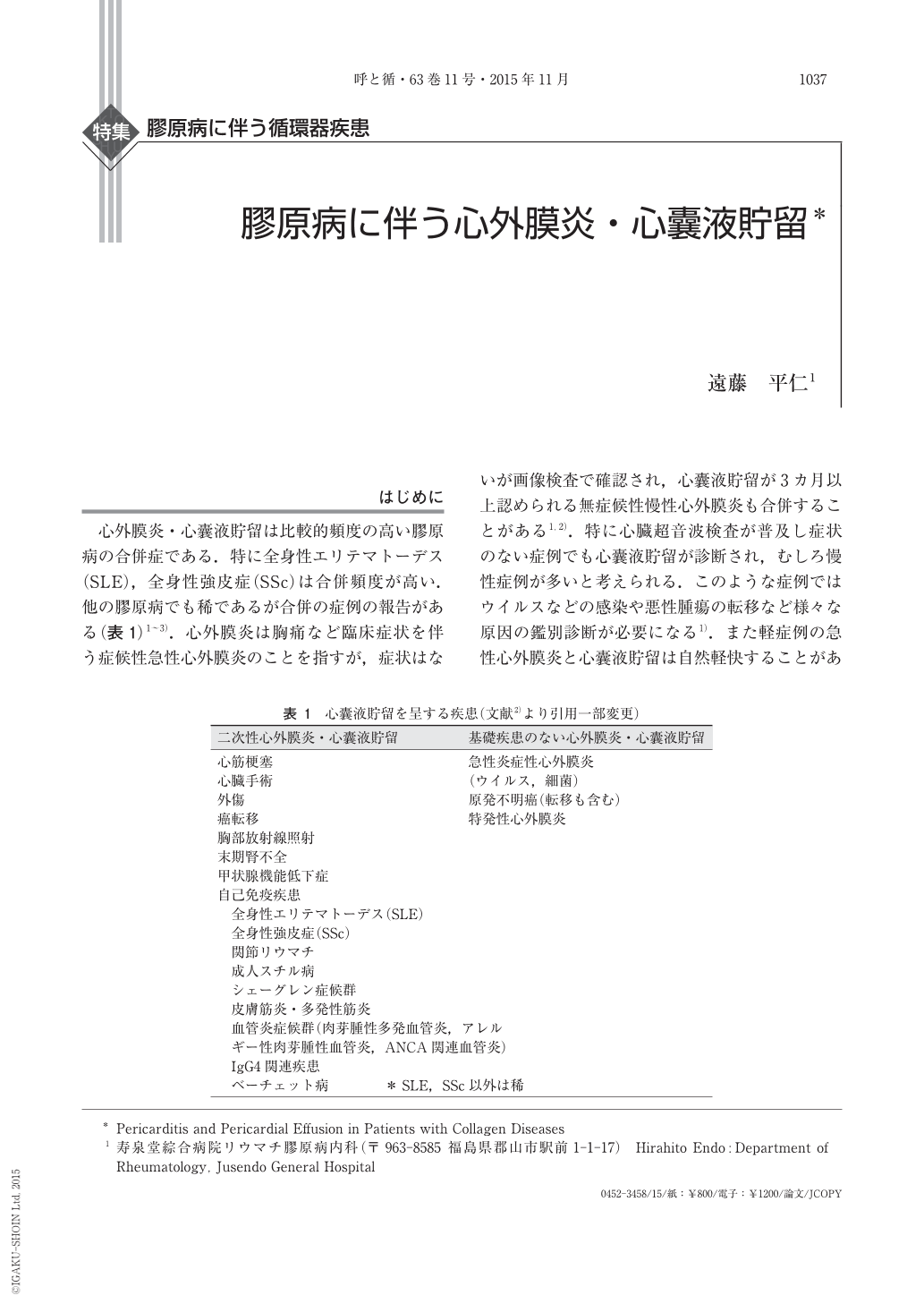
はじめに 心外膜炎・心囊液貯留は比較的頻度の高い膠原病の合併症である.特に全身性エリテマトーデス(SLE),全身性強皮症(SSc)は合併頻度が高い.他の膠原病でも稀であるが合併の症例の報告がある(表1)1〜3).心外膜炎は胸痛など臨床症状を伴う症候性急性心外膜炎のことを指すが,症状はないが画像検査で確認され,心囊液貯留が3カ月以上認められる無症候性慢性心外膜炎も合併することがある1,2).特に心臓超音波検査が普及し症状のない症例でも心囊液貯留が診断され,むしろ慢性症例が多いと考えられる.このような症例ではウイルスなどの感染や悪性腫瘍の転移など様々な原因の鑑別診断が必要になる1).また軽症例の急性心外膜炎と心囊液貯留は自然軽快することがある.欧米の報告では胸痛などの自覚症状があり救急部を受診するのは約5%程度である.死亡率は約1.1%であり心筋炎を併発し重症不整脈や心不全で死亡している2).膠原病に合併した心外膜炎・心囊液貯留は他の病態によるものを除外診断し,各疾患の疾患活動性の評価により治療方針を決める.各膠原病の疾患ごとに胸膜炎の病態形成が異なるため治療,特にステロイド療法の適応について相違点があることに注意する必要がある.特にSScとSLEや他の膠原病合併例は対応が異なる(表2).
1 0 0 0 OA 全身性エリテマトーデスに合併しステロイド大量療法が奏功した骨髄線維症の1例
- 著者
- 吉田 秀 遠藤 平仁 田中 淳一 飯塚 進子 木村 美保 橋本 篤 田中 住明 石川 章 廣畑 俊成 近藤 啓文
- 出版者
- 一般社団法人 日本臨床リウマチ学会
- 雑誌
- 臨床リウマチ (ISSN:09148760)
- 巻号頁・発行日
- vol.20, no.4, pp.302-309, 2008-12-30 (Released:2016-11-30)
- 参考文献数
- 26
A woman of 50 years of age who had a 13-year history of hypothyroidism was diagnosed with systemic lupus erythematosus (SLE) with butterfly rash, leukopenia, positivity of antinuclear antibody, anti-DNA antibody and anti-Sm antibody. Two years later, she developed nephritis (WHO type IV) and remitted with corticosteroid pulse and intermittent intravenous cyclophosphamide pulse therapy (IVCY). Four years after the onset of SLE, she relapsed with proteinuria and leukopenia when she was taking 9 mg/day of prednisolone (PSL) but she stopped all the medication of her own accord. Four months passed without any therapy, she was admitted to our hospital with disturbance of consciousness and anasarca. Laboratory findings showed pancytopenia (WBC 1300/μl, RBC 233×10⁴/μl, Hb6.9g/dl, Plt3.6×10⁴/μl), aggravation of lupus nephritis and hypothyroidism. Chest X-ray and ultrasonography demonstrated pleural and pericardial effusion and the absence of hepatosplenomegaly. She was also diagnosed with myelofibrosis upon bone marrow inspection. Three instances of corticosteroid pulse therapy, oral corticosteroid (PSL was tapered from 50 mg/day) and supplement therapy of levothyroxine improved every symptom and pancytopenia. The second bone marrow biopsy showed reduced fibrosis and recovery of bone marrow cells. These findings implied the secondary myelofibrosis caused by SLE because the myelofibrosis came along with aggravation of SLE and corticosteroid therapy was effective. This is a rare case of SLE in which myelofibrosis improved by high-dose corticosteroid therapy, which was confirmed by bone marrow biopsy and suggests the pathogenic mechanisms for myelofibrosis.
1 0 0 0 OA 炎症収束因子—自然免疫から獲得免疫への橋梁—
- 著者
- 遠藤 平仁
- 出版者
- 日本臨床免疫学会
- 雑誌
- 日本臨床免疫学会会誌 (ISSN:09114300)
- 巻号頁・発行日
- vol.36, no.3, pp.156-161, 2013 (Released:2013-06-30)
- 参考文献数
- 32
- 被引用文献数
- 2 2
体外からの細菌感染や外傷などによる急性炎症は早期に血管浸過性亢進と好中球を中心とする炎症細胞の浸潤が起こる.炎症により破壊された組織は正常の組織に修復される.急性炎症で好中球の浸潤がおこりその後マクロファージ,リンパ球の浸潤と異物の貪食除去の炎症収束,組織修復と生体反応は展開する.急性炎症は自己制御され収束(Resolution)し組織修復する.この転換期に脂質メデイエータのLipoxinやResolvinや抗炎症サイトカインIL-10やアデイポカインChemerinなど多くの因子が作用する.Lipoxinはリポキシゲナーゼ(LOX)により合成される.急性炎症の早期は5-リポキシゲナーゼ(5-LOX)がロイコトリエンB4(LTB4)を合成し好中球の炎症部位に遊走させる.その後マクロファージの15-LOXや血小板の12-LOXが誘導され,5-LOX,と12-LOXまたは15-LOXの2つの酵素を介してLXA4またはLXB4が合成される.このLipoxinは強い抗炎症作用を有している.LXA4はG蛋白結合型受容体ALXに結合し好中球の遊走抑制,マクロファージの遊走活性化などを生じ炎症収束過程の早期に作用する.Chemerinは好中球からのプロテアーゼにより前駆蛋白より活性化されマクロファージ,樹状細胞遊走,抗炎症作用を起こす.またアセチルサリチル酸(アスピリン)の作用したシクロオキシゲナーゼ(COX2)はPGE2産生を抑制し,さらに5-LOXと共に15-epiLXA4を産生し強い抗炎症作用を示す.以上の炎症の収束(Resolution)は自然免疫と獲得免疫を繋ぐ能動的な過程であり新たな炎症治療の戦略の標的である.
