2 0 0 0 OA 口内法X線撮影法の始祖たち
- 著者
- 小林 馨
- 出版者
- 特定非営利活動法人 日本歯科放射線学会
- 雑誌
- 歯科放射線 (ISSN:03899705)
- 巻号頁・発行日
- vol.62, no.1, pp.10-16, 2022 (Released:2022-10-04)
- 参考文献数
- 54
In the present study, we reconsider the pioneers of the intraoral radiography. In intraoral radiography, the bisecting-angle technique was first published by Weston Andrew Valleau Price in 1904, and Antoni Cieszyński devised the same method in 1907. The paralleling technique was first used by Charles Edmund Kells in 1896. Then, in 1911, Franklin W. McCormack introduced the long-distance technique into the paralleling technique and published his findings in 1920. It was integrated and improved, and was popularized by Gordon M. Fitzgerald as a long cone paralleling technique between 1947 and 1950. The author proposes that the above should be the established theory in Japan.
1 0 0 0 OA 咀嚼筋腱・腱膜過形成症のMR画像診断の現状
- 著者
- 小林 馨 下田 信治 依田 哲也 覚道 健治
- 出版者
- 一般社団法人 日本顎関節学会
- 雑誌
- 日本顎関節学会雑誌 (ISSN:09153004)
- 巻号頁・発行日
- vol.21, no.1, pp.35-39, 2009 (Released:2012-02-15)
- 参考文献数
- 8
- 被引用文献数
- 3
1 0 0 0 OA 顎関節部のjoint effusion像と臨床症状との関連 -経時的変化を中心に-
- 著者
- 地挽 雅人 浅田 洸一 豊田 長隆 菊地 健太郎 荒井 智彦 平下 光輝 野上 喜史 石橋 克禮 小林 馨
- 出版者
- 一般社団法人 日本顎関節学会
- 雑誌
- 日本顎関節学会雑誌 (ISSN:09153004)
- 巻号頁・発行日
- vol.9, no.1, pp.60-71, 1997-06-20 (Released:2010-08-06)
- 参考文献数
- 17
- 被引用文献数
- 2
MR画像におけるjoint effusion (以下effusion) と臨床症状との関連, 特に経時的変化について検討した。対象は1993年10月から96年4月までに撮影したMR画像のうちeffusionを認めた33例で, その臨床診断は顎関節症29例, リウマチ性顎関節炎1例, 陳旧性脱臼1例, synovial osteochondromatosis 1例, 下顎骨周囲炎に後遺する下顎頭萎縮1例であった。男性5例, 女性28例, 年齢は14-65歳, 平均38.9歳であった。effusionは矢状面SE法MR撮像T2画像で上または下関節腔に, 高信号を示し, かつT1画像で低信号を示すものとした。臨床症状, X線写真所見, 円板動態との関連と, このうち16例20関節は2か月から1年9か月後に経時的変化について検討した。その結果, 初回MR所見でeffusionを認めた部位は上関節腔37, 下関節腔2関節で, 溝口らの分類によると帯状17, 太線状7, 線状7, 点状8関節であった。主訴は顎関節部の疼痛が多かった。また復位を伴わない円板前方転位例に多く, 特に帯状の例にその傾向が強かった。開口度は16-54mm, 平均37.3mmであった。X線写真所見で骨変形を認めた例にやや多いが, 約半数の関節では骨構成体に変化はなかった。経時的にはeffusionが消失したもの7関節, 大きさが縮小したもの5関節, 変化のないもの7関節, 性状が変化しているもの1関節であった。この像は下顎頭が, 滑膜表面に大きな応力が加わることによって, 滑膜や関節構成組織より, 組織液や何らかの融解産物が滑液中に浸出した結果と考えられた。
1 0 0 0 Square-Mandible 顔貌を伴う開口制限の1例
1 0 0 0 OA 体格指数と舌筋の脂肪化が無呼吸・低呼吸指数に及ぼす影響
- 著者
- 玉井 和樹 小林 馨 五十嵐 千浪 小佐野 貴識
- 出版者
- 特定非営利活動法人 日本歯科放射線学会
- 雑誌
- 歯科放射線 (ISSN:03899705)
- 巻号頁・発行日
- vol.53, no.3, pp.21-27, 2013 (Released:2013-12-10)
- 参考文献数
- 23
Objective: In recent years, it has been postulated that a change in muscle function is associated with the etiology of obstructive sleep apnea (OSA). Saito and colleagues previously reported the impact of obesity on the properties of the lingual muscles (genioglossus and geniohyoid) in rats (Arch Oral Biol (2010; 55(10): 803-808)). However, in previous images, fat to muscle metamorphosis was not shown in humans. Here, we provide evidence of fatty metamorphosis in the lingual muscles using computed tomography (CT) images of patients with suspected OSA.Materials and methods: The subjects were 62 patients (47 male, 15 female) with suspected OSA, who visited Tsurumi University School of Dental Medicine from November 2007 to October 2011. All subjects gave informed consent to take part in the study. The subjects underwent CT evaluations at the image diagnosis department of the hospital. Sex, age, body mass index (BMI), and apnea hypopnea index (AHI) were recorded for each patient. Inferior airway space and the total value of length and width of the inferior airway space (TIAS) were also measured. The degree of fat to muscle metamorphosis was measured using CT. Aze Win image analysis software was used to set a region of interest (ROI) of 30mm2 on the belly muscle of the lingual muscles. We measured CT levels of four ROIs (both sides of the central area and both sides of the posterior area) in the genioglossus muscles and two sizes of ROI (both sides of the central area) in geniohyoid muscles. Values were quantified and compared statistically.Results: The median values (25% quartile deviation; 75% quartile deviation) of the patient's age, BMI (kg/m2), AHI, genioglossus CT level, geniohyoid muscle CT level, and TIAS were 51.50 years old (42.75, 62.25), 24.00 (22.00, 26.00), 24.35 (11.40, 36.10), 123.05 (90.95, 135.70), 111.20 (104.80, 116.30), and 34.65 (25.97, 40.62), respectively. The results of a multiple regression model were analyzed using Amos (Ver. 6) software where the standardized estimates of the BMI were -0.50 (p = 0.000) for the genioglossus muscle and -0.42 (p = 0.000) for the geniohyoid muscle. The standardized estimate of BMI of the distance of the TIAS was -0.55 (p = 0.001). The standardized estimate of AHI of the TIAS was -0.48 (p = 0.000).Conclusion: Consistent with the report of Saito et al., we showed evidence of fatty metamorphosis of lingual muscles of humans with effects of the TIAS and AHI.
1 0 0 0 Square-Mandible顔貌を伴う開口制限の1例
- 著者
- 岡田 成生 上野 泰宏 星 健太郎 伊藤 弘人 神部 芳則 草間 幹夫 小林 馨
- 出版者
- 特定非営利活動法人 日本歯科放射線学会
- 雑誌
- 歯科放射線 (ISSN:03899705)
- 巻号頁・発行日
- vol.48, no.1, pp.8-11, 2008
We report a patient with jaw trismus demonstrating Square-Mandible face.<BR>Clinically, the patient had a square face with prominent masseter muscles and mandibular angles. The interincisal opening was 26mm, with limited lateral and anteroposterior mobility.<BR>The clinical diagnosis was restricted mouth opening due to bilateral hyperplasia of the masseter muscle aponeuorses. Masseter muscle myotomy with aponeurectomy was performed bilaterally by an intraoral approach. Mouth opening improved to 40mm after incision of the aponeuroses.<BR>An examination of the patient one year after surgery demonstrated an interincisal opening of 37mm without strain.
- 著者
- 井口 貞彦 岩村 浩幸 西崎 稔 林 昭夫 瀬の口 和彦 小林 馨 坂木 克人 蜂谷 勝敏 市岡 由美子 川村 雅範
- 出版者
- The Pharmaceutical Society of Japan
- 雑誌
- Chemical and Pharmaceutical Bulletin (ISSN:00092363)
- 巻号頁・発行日
- vol.40, no.6, pp.1462-1469, 1992-06-25 (Released:2008-03-31)
- 参考文献数
- 12
- 被引用文献数
- 115 152
A novel, highly cardioselective ultra short-acting β-blocker, ONO-1101, has been developed for application in the emergency treatment of tachycardia and better control of heart rate in surgery. This agent is approximately nine times more potent in β-blocking activity in vivo and eight times more cardioselective in vitro than esmolol. This β-blocking drug has a short duration of activity, enabling rapid recovery after cessation of administration if side effects occur. It can be used safely in patients suffering from acute heart disease and represents a major therapeutic advance in the treatment of heart disease.
1 0 0 0 歯科放射線学
- 著者
- 岡野友宏 小林馨 有地榮一郎編 浅海淳一 [ほか] 執筆
- 出版者
- 医歯薬出版
- 巻号頁・発行日
- 2013
- 著者
- 小林 馨
- 出版者
- 明治大学史学地理学会
- 雑誌
- 駿台史學 = Sundai historical review : the journal of the Historico-Geographical Association of Meiji University (ISSN:05625955)
- 巻号頁・発行日
- no.152, pp.25-51, 2014-09
1 0 0 0 口腔外科の疾患と治療
- 著者
- 栗田賢一 覚道健治 小林馨編
- 出版者
- 永末書店
- 巻号頁・発行日
- 1998
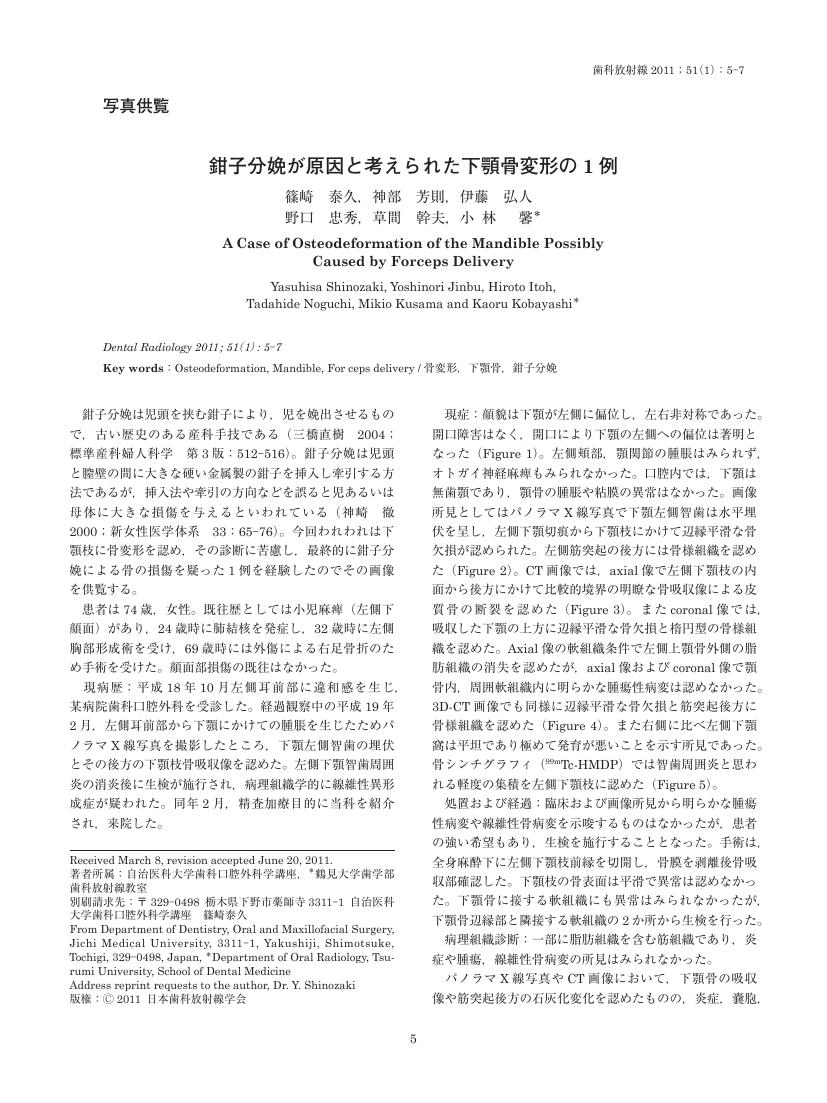
1 0 0 0 OA 鉗子分娩が原因と考えられた下顎骨変形の1例
- 著者
- 篠崎 泰久 神部 芳則 伊藤 弘人 野口 忠秀 草間 幹夫 小林 馨
- 出版者
- 特定非営利活動法人 日本歯科放射線学会
- 雑誌
- 歯科放射線 (ISSN:03899705)
- 巻号頁・発行日
- vol.51, no.1, pp.5-7, 2011 (Released:2011-08-05)
1 0 0 0 歯科用CTによる下顎管と下顎智歯の位置関係の観察
- 著者
- 橋爪 敦子 中川 洋一 石井 久子 小林 馨
- 出版者
- Japanese Society of Oral and Maxillofacial Surgeons
- 雑誌
- 日本口腔外科学会雑誌 (ISSN:00215163)
- 巻号頁・発行日
- vol.50, no.1, pp.1-10, 2004-01-20
- 参考文献数
- 19
- 被引用文献数
- 7 3
We describe the preoperative use of limited cone beam computed tomography (CT) with a dental CT scanner for the assessment of mandibular third molars before extraction. Cone beam CT provides 42.7-mm-high and 30-mm-wide rectangular solid images, with a resolution of less than 0.2mm.<BR>The positional relationship between the mandibular third molars and the mandibular canal was examined by dental CT. Sixty-eight lower third molars of 62 patients whose teeth were superimposed on the mandibular canal on periapical or panoramic radiographs were studied. Dental CT scans clearly demonstrated the positional relationship between the mandibular canal and the teeth. The mandibular canal was located buccally to the roots of 16 teeth, lingually to the roots of 27 teeth, inferiorly to the roots of 23 teeth, and between the roots of 2 teeth. The presence of bone between the mandibular canal and the teeth was not noted in 7 of 16 buccal cases, 24 of 27 lingual cases, and 10 of 23 inferior cases on dental CT scans, suggesting that the cana was in contact with the teeth.<BR>Fifty-nine of the 68 mandibular third molars were surgically removed, and postoperative transient hypoesthesia occurred in 4 patients. Dental CT scans showed no bone between the mandibular canal and the teeth in all 4 patients. Hypoesthesia was not related to the bucco-lingual location of the mandibular canal or to the extent of bone loss between the canal and the teeth. However, hypoesthesia did not occur in patients with bone between the mandibular canal and the teeth. Thus, information on the distance between the canal and teeth on dental CT scans was useful for predicting the risk of inferior alveolar nerve damage.<BR>Because of its high resolution and low radiation dose, cone beam CT was useful for examination before mandibular third molar surgery.


