11 0 0 0 OA JCS 2022 Guideline Focused Update on Diagnosis and Treatment in Patients With Stable Coronary Artery Disease
- 著者
- Shintaro Nakano Shun Kohsaka Taishiro Chikamori Kenji Fukushima Yoshio Kobayashi Ken Kozuma Susumu Manabe Hitoshi Matsuo Masato Nakamura Takayuki Ohno Mitsuaki Sawano Koichi Toda Yasunori Ueda Hiroyoshi Yokoi Yodo Gatate Tokuo Kasai Yoshiaki Kawase Naoya Matsumoto Hitoshi Mori Ryo Nakazato Nozomi Niimi Yuichi Saito Ayumi Shintani Ippei Watanabe Yusuke Watanabe Yuji Ikari Masahiro Jinzaki Masami Kosuge Kenichi Nakajima Takeshi Kimura on behalf of the JCS Joint Working Group
- 出版者
- The Japanese Circulation Society
- 雑誌
- Circulation Journal (ISSN:13469843)
- 巻号頁・発行日
- pp.CJ-21-1041, (Released:2022-03-11)
- 参考文献数
- 245
- 被引用文献数
- 40
- 著者
- Masakazu Yamagishi Nagara Tamaki Takashi Akasaka Takanori Ikeda Kenji Ueshima Shiro Uemura Yutaka Otsuji Yasuki Kihara Kazuo Kimura Takeshi Kimura Yoshiki Kusama Shinichiro Kumita Hajime Sakuma Masahiro Jinzaki Hiroyuki Daida Yasuchika Takeishi Hiroshi Tada Taishiro Chikamori Kenichi Tsujita Kunihiko Teraoka Kenichi Nakajima Tomoaki Nakata Satoshi Nakatani Akihiko Nogami Koichi Node Atsushi Nohara Atsushi Hirayama Nobusada Funabashi Masaru Miura Teruhito Mochizuki Hiroyoshi Yokoi Kunihiro Yoshioka Masafumi Watanabe Toshihiko Asanuma Yuichi Ishikawa Takahiro Ohara Koichi Kaikita Tokuo Kasai Eri Kato Hiroshi Kamiyama Masaaki Kawashiri Keisuke Kiso Kakuya Kitagawa Teruhito Kido Toshio Kinoshita Tomonari Kiriyama Teruyoshi Kume Akira Kurata Satoshi Kurisu Masami Kosuge Eitaro Kodani Akira Sato Yasutsugu Shiono Hiroki Shiomi Junichi Taki Masaaki Takeuchi Atsushi Tanaka Nobuhiro Tanaka Ryoichi Tanaka Takuya Nakahashi Takehiro Nakahara Akihiro Nomura Akiyoshi Hashimoto Kenshi Hayashi Masahiro Higashi Takafumi Hiro Daisuke Fukamachi Hitoshi Matsuo Naoya Matsumoto Katsumi Miyauchi Masao Miyagawa Yoshitake Yamada Keiichiro Yoshinaga Hideki Wada Tetsu Watanabe Yukio Ozaki Shun Kohsaka Wataru Shimizu Satoshi Yasuda Hideaki Yoshino on behalf of the Japanese Circulation Society Working Group
- 出版者
- The Japanese Circulation Society
- 雑誌
- Circulation Journal (ISSN:13469843)
- 巻号頁・発行日
- pp.CJ-19-1131, (Released:2021-02-16)
- 参考文献数
- 1401
- 被引用文献数
- 50
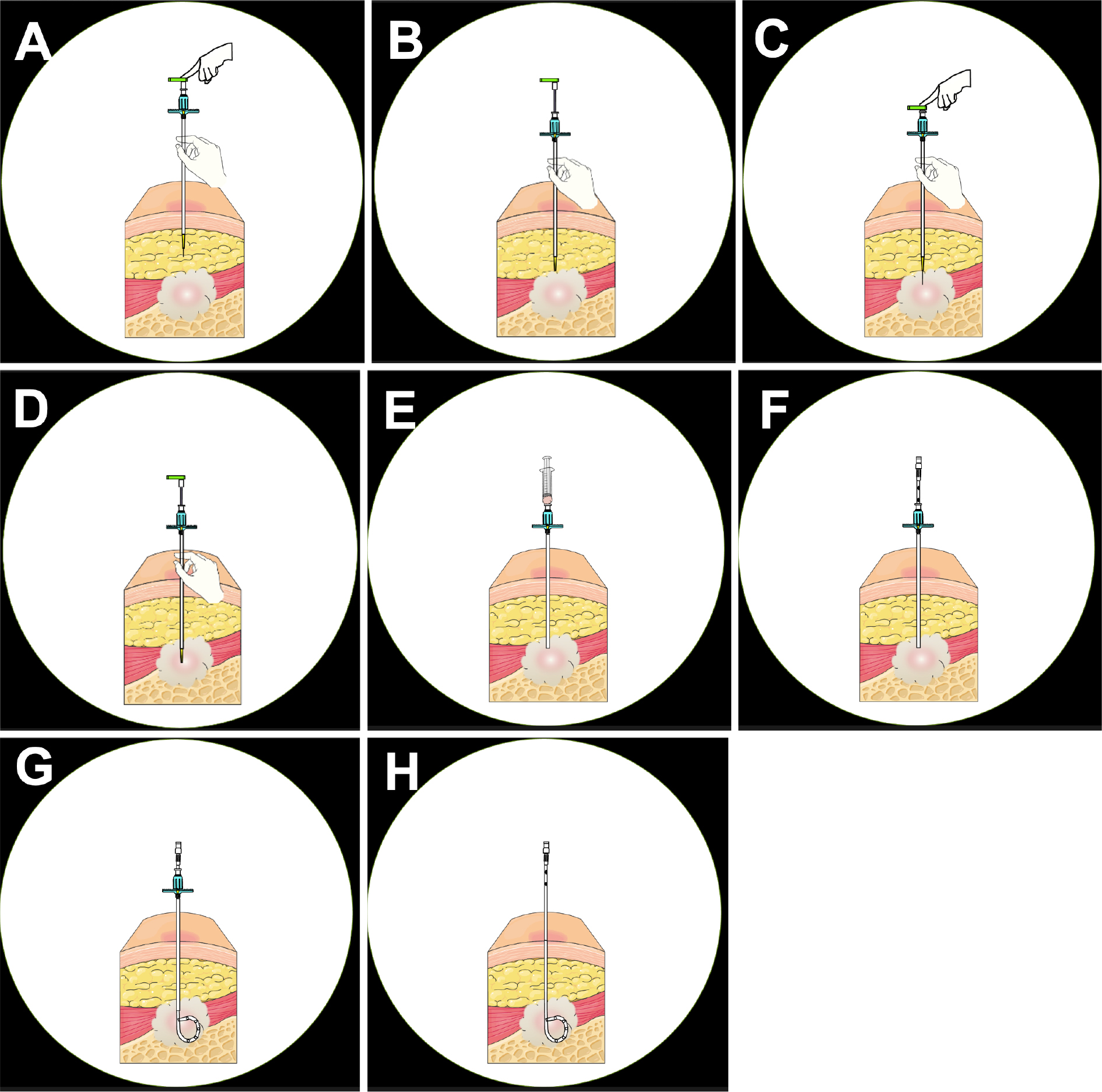
2 0 0 0 OA Computed Tomography-guided Drainage with Modified Trocar Technique Using a Drainaway Drainage Kit
- 著者
- Koji Togawa Seishi Nakatsuka Jitsuro Tsukada Nobutake Ito Yosuke Yamamoto Togo Kogo Hiroki Yoshikawa Manabu Misu Masashi Tamura Shigeyoshi Soga Masanori Inoue Hideki Yashiro Tadayoshi Kurata Masahiro Okada Masahiro Jinzaki
- 出版者
- Japanese Society of Interventional Radiology
- 雑誌
- Interventional Radiology (ISSN:24320935)
- 巻号頁・発行日
- vol.8, no.3, pp.130-135, 2023-11-01 (Released:2023-11-01)
- 参考文献数
- 10
Purpose: Image-guided percutaneous drainage for abscesses is known as a safe and effective treatment. The computed tomography-guided percutaneous drainage kit Drainaway (SB Kawasumi Co., Ltd.), developed on the basis of a modified trocar method, has made it possible to complete the procedure only under computed tomography guidance without radiographic fluoroscopy. This study investigated the feasibility and safety of Drainaway for abscess drainage.Material and Methods: In this retrospective observational study, 28 procedures in 27 patients (18 men and 9 women; age 67.0 ± 12.3 years) who underwent computed tomography-guided drainage using Drainaway between March and December 2021 at seven affiliated hospitals were analyzed. Patients with symptomatic, puncturable on computed tomography and refractory abscesses were included. Technical success (successful drainage with computed tomography alone), primary clinical success (successful drainage with Drainaway alone), secondary clinical success (avoidance of surgery), and complications were evaluated.Results: The sites of the abscesses were the intraperitoneal, retroperitoneal, and thoracic cavities in 19, 5, and 2 patients, respectively, and subcutaneous tissue in 1 patient. The mean size of the abscesses was 7.1 ± 3.4 cm. The technical success rate was 96.4%; the ligament of the puncture route could not be penetrated in one case. The primary clinical success rate was 77.8%, whereas the secondary clinical success rate of catheter upsizing or replacement was 96.3%. Complications included one case of biliary pleurisy that required drainage.Conclusions: Drainaway is a useful device that allows abscess drainage using only computed tomography guidance without radiographic fluoroscopy.
- 著者
- Shintaro Nakano Shun Kohsaka Taishiro Chikamori Kenji Fukushima Yoshio Kobayashi Ken Kozuma Susumu Manabe Hitoshi Matsuo Masato Nakamura Takayuki Ohno Mitsuaki Sawano Koichi Toda Yasunori Ueda Hiroyoshi Yokoi Yodo Gatate Tokuo Kasai Yoshiaki Kawase Naoya Matsumoto Hitoshi Mori Ryo Nakazato Nozomi Niimi Yuichi Saito Ayumi Shintani Ippei Watanabe Yusuke Watanabe Yuji Ikari Masahiro Jinzaki Masami Kosuge Kenichi Nakajima Takeshi Kimura on behalf of the JCS Joint Working Group
- 出版者
- The Japanese Circulation Society
- 雑誌
- Circulation Journal (ISSN:13469843)
- 巻号頁・発行日
- vol.86, no.5, pp.882-915, 2022-04-25 (Released:2022-04-25)
- 参考文献数
- 245
- 被引用文献数
- 40
1 0 0 0 OA A Versatile MR Elastography Research Tool with a Modified Motion Signal-to-noise Ratio Approach
- 著者
- Daiki Ito Tetsushi Habe Tomokazu Numano Shigeo Okuda Shigeyoshi Soga Masahiro Jinzaki
- 出版者
- Japanese Society for Magnetic Resonance in Medicine
- 雑誌
- Magnetic Resonance in Medical Sciences (ISSN:13473182)
- 巻号頁・発行日
- pp.mp.2022-0149, (Released:2023-04-12)
- 参考文献数
- 41
Purpose: This study aimed to facilitate research progress in MR elastography (MRE) by providing a versatile and convenient application for MRE reconstruction, namely the MRE research tool (MRE-rTool). It can be used for a series of MRE image analyses, including phase unwrapping, arbitrary bandpass and directional filtering, noise assessment of the wave propagation image (motion SNR), and reconstruction of the elastogram in both 2D and 3D MRE acquisitions. To reinforce the versatility of MRE-rTool, the conventional method of motion SNR was modified into a new method that reflects the effects of image filtering.Methods: MRE tests of the phantom and liver were performed using different estimation algorithms for stiffness value (algebraic inversion of the differential equation [AIDE], local frequency estimation [LFE] in MRE-rTool, and multimodel direct inversion [MMDI] in clinical reconstruction) and acquiring dimensions (2D and 3D acquisitions). This study also tested the accuracy of masking low SNR regions using modified and conventional motion SNR under various mechanical vibration powers.Results: The stiffness values estimated using AIDE/LFE in MRE-rTool were comparable to that of MMDI (phantom, 3.71 ± 0.74, 3.60 ± 0.32, and 3.60 ± 0.54 kPa in AIDE, LFE, and MMDI; liver, 2.26 ± 0.31, 2.74 ± 0.16, and 2.21 ± 0.26 kPa in AIDE, LFE, and MMDI). The stiffness value in 3D acquisition was independent of the direction of the motion-encoding gradient and was more accurate than that of 2D acquisition. The masking of low SNR regions using the modified motion SNR worked better than that in the conventional motion SNR for each vibration power, especially when using a directional filter.Conclusion: The performance of MRE-rTool on test data reached the level required in clinical MRE studies. MRE-rTool has the potential to facilitate MRE research, contribute to the future development of MRE, and has been freely released online.
- 著者
- Ho Lee Tatsuya Suzuki Yohei Okada Hiromu Tanaka Satoshi Okamori Hirofumi Kamata Makoto Ishii Masahiro Jinzaki Koichi Fukunaga
- 出版者
- The Keio Journal of Medicine
- 雑誌
- The Keio Journal of Medicine (ISSN:00229717)
- 巻号頁・発行日
- pp.2021-0012-OA, (Released:2021-11-11)
- 参考文献数
- 23
- 被引用文献数
- 2
Coronavirus disease 2019 (COVID-19) was first reported in Wuhan, China, in December 2019 as an outbreak of pneumonia of unknown origin. Previous studies have suggested the utility of chest computed tomography (CT) in the diagnosis of COVID-19 because of its high sensitivity (93%–97%), relatively simple procedure, and rapid test results. This study, performed in Japan early in the epidemic when COVID-19 prevalence was low, evaluated the diagnostic accuracy of chest CT in a population presenting with lung diseases having CT findings similar to those of COVID-19. We retrospectively included all consecutive patients (≥18 years old) presenting to the outpatient department of Keio University Hospital between March 1 and May 31, 2020, with fever and respiratory symptoms. We evaluated the performance of diagnostic CT for COVID-19 by using polymerase chain reaction (PCR) results as the reference standard. We determined the numbers of false-positive (FP) results and assessed the clinical utility using decision curve analysis. Of the 175 patients, 22 were PCR-positive. CT had a sensitivity of 68% and a specificity of 57%. Patients with FP results on CT diagnosis were mainly diagnosed with diseases mimicking COVID-19, e.g., interstitial lung disease. Decision curve analysis indicated that the clinical utility of CT imaging was limited. The diagnostic performance of CT for COVID-19 was inadequate in an area with low COVID-19 prevalence and a high prevalence of other lung diseases with chest CT findings similar to those of COVID-19. Considering this insufficient diagnostic performance, CT findings should be evaluated in the context of additional medical information to diagnose COVID-19.
- 著者
- Toru Miyoshi Kazuhiro Osawa Hiroshi Ito Susumu Kanazawa Takeshi Kimura Hiroki Shiomi Sachio Kuribayashi Masahiro Jinzaki Akio Kawamura Hiram Bezerra Stephan Achenbach Bjarne L. Nørgaard
- 出版者
- 日本循環器学会
- 雑誌
- Circulation Journal (ISSN:13469843)
- 巻号頁・発行日
- vol.79, no.2, pp.406-412, 2015-01-23 (Released:2015-01-23)
- 参考文献数
- 29
- 被引用文献数
- 5 23
Background:Recently, a non-invasive method using computational fluid dynamics to calculate vessel-specific fractional flow reserve (FFRCT) from routinely acquired coronary computed tomography angiography (CTA) was described. The Analysis of Coronary Blood Flow Using CT Angiography: Next Steps (NXT) trial, which was a prospective, multicenter trial including 254 patients with suspected coronary artery disease, noted high diagnostic performance of FFRCTcompared with invasive FFR. The aim of this post-hoc analysis was to assess the diagnostic performance of non-invasive FFRCTvs. standard stenosis quantification on coronary CTA in the Japanese subset of the NXT trial.Methods and Results:A total of 57 Japanese participants were included from Okayama University (n=36), Kyoto University (n=17), and Keio University (n=4) Hospitals. Per-patient diagnostic accuracy of FFRCT(74%; 95% confidence interval [CI]: 60–85%) was higher than for coronary CTA (47%; 95% CI: 34–61%, P<0.001) arising from improved specificity (63% vs. 27%, P<0.001). FFRCTcorrectly reclassified 53% of patients and 63% of vessels with coronary CTA false positives as true negatives. When patients with Agatston score >1,000 were excluded, per-patient accuracy of FFRCTwas 83% with a high specificity of 76%, similar to the overall NXT trial findings.Conclusions:FFRCThas high diagnostic performance compared with invasive FFR in the Japanese subset of patients in the NXT trial. (Circ J 2015; 79: 406–412)
- 著者
- Masahiro Jinzaki Kozo Sato Yutaka Tanami Minoru Yamada Sachio Kuribayashi Toshihisa Anzai Yasushi Asakura Satoshi Ogawa
- 出版者
- The Japanese Circulation Society
- 雑誌
- Circulation Journal (ISSN:13469843)
- 巻号頁・発行日
- vol.70, no.12, pp.1661-1662, 2006 (Released:2006-11-25)
- 参考文献数
- 7
- 被引用文献数
- 23 24
Background A method of displaying coronary computed tomography (CT) angiography, which enables evaluation of coronary artery disease (CAD) with fewer images and is understandable to the third person, is preferable. Methods and Results A maximum intensity projection image was created in which contrast media in the ventricles is eliminated, enabling an overview of CAD in a single 3-dimensional (D) image that can be rotated to be viewed at various angles and is easily understood by a third person. Conclusions A novel method of displaying coronary CT angiography in a single 3-D image has been developed and we believe it should become available for many workstations. (Circ J 2006; 70: 1661 - 1662)

