- 著者
- 田中 政義 荒川 佳也
- 出版者
- 公益社団法人 日本放射線技術学会
- 雑誌
- 日本放射線技術学会雑誌 (ISSN:03694305)
- 巻号頁・発行日
- vol.36, no.5, pp.717-718, 1980
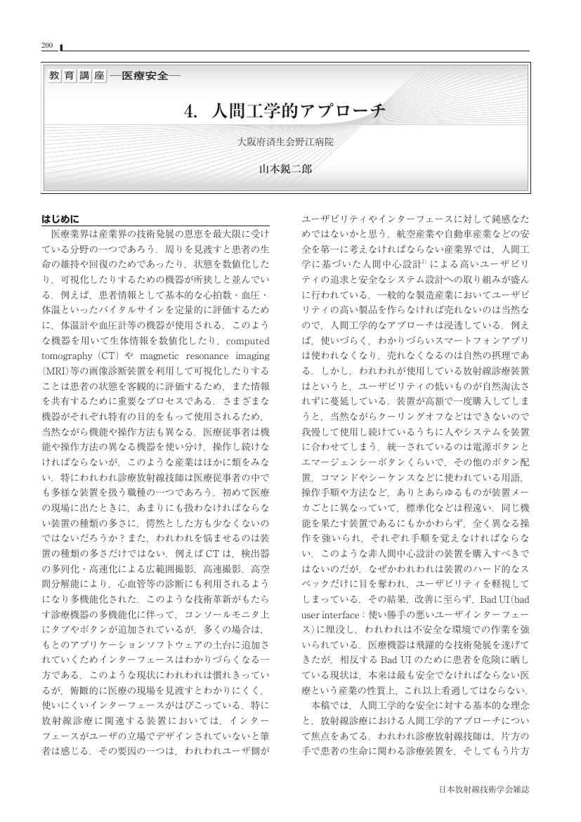
1 0 0 0 4. 人間工学的アプローチ
- 著者
- 山本 鋭二郎
- 出版者
- 公益社団法人 日本放射線技術学会
- 雑誌
- 日本放射線技術学会雑誌 (ISSN:03694305)
- 巻号頁・発行日
- vol.77, no.2, pp.200-211, 2021 (Released:2021-02-20)
- 参考文献数
- 13
- 著者
- 荒井 博史 久保 直樹 表 英彦 高橋 典子 勝浦 秀則 鈴木 幸太郎 古館 正従
- 出版者
- 公益社団法人 日本放射線技術学会
- 雑誌
- 日本放射線技術学会雑誌 (ISSN:03694305)
- 巻号頁・発行日
- vol.44, no.7, 1988
1 0 0 0 2 多目的カセッテホルダについて
- 著者
- 宮原 清英 岩田 康平
- 出版者
- 公益社団法人 日本放射線技術学会
- 雑誌
- 日本放射線技術学会雑誌 (ISSN:03694305)
- 巻号頁・発行日
- vol.54, no.9, 1998
1 0 0 0 エリアモニタ監視方式による感染症医療廃棄物の管理
- 著者
- 柳沢 正道 丸 繁勘 椎葉 眞一
- 出版者
- 公益社団法人 日本放射線技術学会
- 雑誌
- 日本放射線技術学会雑誌 (ISSN:03694305)
- 巻号頁・発行日
- vol.58, no.1, pp.130-132, 2002
- 参考文献数
- 4
- 被引用文献数
- 1 1
- 著者
- 笹本 和夫 長山 忠雄
- 出版者
- 公益社団法人 日本放射線技術学会
- 雑誌
- 日本放射線技術学会雑誌 (ISSN:03694305)
- 巻号頁・発行日
- vol.44, no.8, pp.1034, 1988-08-01 (Released:2017-06-28)
1 0 0 0 2.MRI の Radiomics
- 著者
- 酒井 晃二
- 出版者
- 公益社団法人 日本放射線技術学会
- 雑誌
- 日本放射線技術学会雑誌 (ISSN:03694305)
- 巻号頁・発行日
- vol.77, no.8, pp.866-875, 2021 (Released:2021-08-20)
- 参考文献数
- 103
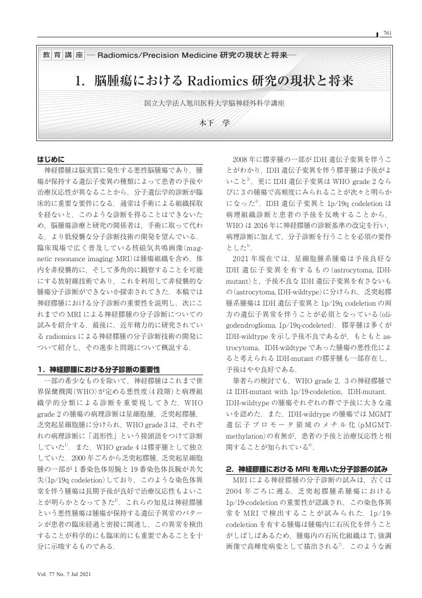
1 0 0 0 1.脳腫瘍における Radiomics 研究の現状と将来
- 著者
- 木下 学
- 出版者
- 公益社団法人 日本放射線技術学会
- 雑誌
- 日本放射線技術学会雑誌 (ISSN:03694305)
- 巻号頁・発行日
- vol.77, no.7, pp.761-764, 2021 (Released:2021-07-20)
- 参考文献数
- 16
- 著者
- 村松 千左子
- 出版者
- 公益社団法人 日本放射線技術学会
- 雑誌
- 日本放射線技術学会雑誌 (ISSN:03694305)
- 巻号頁・発行日
- vol.77, no.7, pp.760, 2021 (Released:2021-07-20)
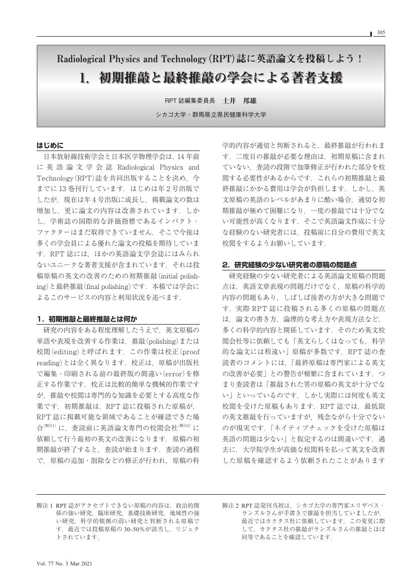
1 0 0 0 1. 初期推敲と最終推敲の学会による著者支援
- 著者
- 土井 邦雄
- 出版者
- 公益社団法人 日本放射線技術学会
- 雑誌
- 日本放射線技術学会雑誌 (ISSN:03694305)
- 巻号頁・発行日
- vol.77, no.3, pp.305-308, 2021 (Released:2021-03-20)
- 参考文献数
- 9
- 著者
- 菅原 時美
- 出版者
- 公益社団法人 日本放射線技術学会
- 雑誌
- 日本放射線技術学会雑誌 (ISSN:03694305)
- 巻号頁・発行日
- vol.21, no.4, 1966
- 著者
- 山城 晶弘 神谷 直紀 大塚 薫 駒津 和浩 伊東 洋一 久保田 展聡 小林 正人
- 出版者
- 公益社団法人 日本放射線技術学会
- 雑誌
- 日本放射線技術学会雑誌 (ISSN:03694305)
- 巻号頁・発行日
- vol.69, no.2, pp.163-169, 2013-02-20 (Released:2013-03-01)
- 参考文献数
- 13
- 被引用文献数
- 2
In magnetic resonance imaging (MRI), the ideal phantom should have similar T1 and T2 values to those of organs of interest for measuring the change in signal intensity, contrast ratio and contrast noise ratio. There have been several reports to develop such a phantom using materials with limited availability or complex methods. In this study, we have developed a simple phantom using indigestible dextrin and soluble calcium at 1.5-tesla MRI. The T1 and T2 values have been reduced by dissolving indigestible dextrin and soluble calcium in distilled water. The similar T1 and T2 values to those of organs (i.e., kidney cortex, kidney medulla, liver, spleen, pancreas, bone marrow, uterus myometrium, uterus endometrium, uterus cervix, prostate, brain white matter, and brain gray matter) have been obtained by varying the concentration of indigestible dextrin and soluble calcium. This phantom is easy to develop and has a potential to increase the accuracy of MRI phantom experiments.
1 0 0 0 OA 肝悪性腫瘍切除術前3DCTにおける門脈相撮影タイミングの最適化
- 著者
- 原田 耕平 千葉 彩佳 溝延 数房 沼澤 香夏子 今井 達也
- 出版者
- 公益社団法人 日本放射線技術学会
- 雑誌
- 日本放射線技術学会雑誌 (ISSN:03694305)
- 巻号頁・発行日
- vol.72, no.11, pp.1098-1104, 2016 (Released:2016-11-20)
- 参考文献数
- 12
- 被引用文献数
- 1
Preoperative three-dimensional computed tomography (3DCT) of the liver is the most important examination in performing preoperative simulation. Detailed visualization of the portal vein using the workstation is critical to enable accurate liver segmentation. However, the timing of imaging in the portal venous phase has mostly been reported equivalent to that of the liver screening examinations commonly performed. The purpose of this study was to examine the optimal timing of image capture to create the best portal vein visualization in preoperative 3DCT of the liver. Seventy-nine patients who underwent hepatectomy for malignant liver tumors were enrolled in this study. All patients were preoperatively examined using protocol A (imaging method separated into a portal venous phase and a hepatic venous phase) and then examined 1 week after surgery using protocol B (normal liver screening protocol). We first established the regions of interest in the portal vein and the hepatic vein and then compared CT values for these regions under protocol A and protocol B. The average CT value of the portal vein in protocol A and B was 239.8±28.1 HU and 202.2±18.5 HU, respectively. The average CT value of the portal vein in protocol A was significantly higher compared with protocol B (p<0.01). By introducing separate timing for portal venous phase imaging before preoperative 3DCT (protocol A), it is possible to satisfactorily depict the portal vein.
1 0 0 0 44. HALF BEAMを用いた腹部単純立位撮影 第二報
- 著者
- 田淵 真弘 松尾 孝人 小橋 義範 水内 敬枝
- 出版者
- 公益社団法人 日本放射線技術学会
- 雑誌
- 日本放射線技術学会雑誌 (ISSN:03694305)
- 巻号頁・発行日
- vol.51, no.3, 1995
- 著者
- 田淵 真弘 松尾 孝人 小橋 義範 水内 敬枝
- 出版者
- 公益社団法人 日本放射線技術学会
- 雑誌
- 日本放射線技術学会雑誌 (ISSN:03694305)
- 巻号頁・発行日
- vol.50, no.10, 1994
1 0 0 0 OA インパルス応答を利用した核磁気共鳴画像のスライス厚測定
- 著者
- 山口 功 石森 佳幸 藤原 康博 谷内田 拓也 吉岡 千絵
- 出版者
- 公益社団法人 日本放射線技術学会
- 雑誌
- 日本放射線技術学会雑誌 (ISSN:03694305)
- 巻号頁・発行日
- vol.68, no.10, pp.1295-1306, 2012-10-20 (Released:2012-10-22)
- 参考文献数
- 13
- 被引用文献数
- 1 1
The purpose of this study was to verify the applicability of measurement of slice thickness of magnetic resonance imaging (MRI) by the delta method, and to discuss the measurement precision by the disk diameter and baseline setup of the slice profile of the delta method. The delta method used the phantom which put in the disk made of acrylic plastic. The delta method measured the full width at half maximum (FWHM) and the full width at tenth maximum (FWTM) from the slice profile of the disk signal. Evaluation of the measurement precision of the delta method by the disk diameter and baseline setup were verified by comparison of the FWHM and FWTM. In addition, evaluation of the applicability of the delta method was verified by comparison of the FWHM and FWTM using the wedge method. The baseline setup had the proper signal intensity of an average of 10 slices in the disk images. There were statistically significant difference in the FWHM between disk diameter of 10 mm and disk diameter of 30 mm and 5 mm. The FWHM of the disk diameter of 10 mm was smaller than the disk diameter of 30 mm and 5 mm. There was no statistically significant difference in the FWHM between the delta method and the wedge method. There is no difference in the effective slice thickness of the delta method and the wedge method. The delta method has an advantage in measurement of thin slice thickness.
1 0 0 0 OA 傾斜板法を用いた3D撮像のスライスプロファイル計測に対する一考察
- 著者
- 吉田 礼 町田 好男 小倉 隆英 田村 元 引地 健生 森 一生
- 出版者
- 公益社団法人 日本放射線技術学会
- 雑誌
- 日本放射線技術学会雑誌 (ISSN:03694305)
- 巻号頁・発行日
- vol.68, no.11, pp.1456-1466, 2012-11-20 (Released:2012-11-21)
- 参考文献数
- 16
- 被引用文献数
- 3 4
Recent progress in variable-flip-angle fast spin-echo technology has further extended the utility of three-dimensional (3D) magnetic resonance imaging (MRI) for clinical application. The slice profile in 3D MRI is the point spread function that has a sync form in principle, whereas a slice profile in 2D imaging provides information on characteristics of selective radio frequency excitation. We investigated the optimal condition to measure 3D slice profiles using a crossed thin-ramps phantom. We found that the profile data should cover a large area in order to evaluate both the main lobe and side lobes in the slice profile, and that the appropriate slice thickness was 2 mm. We also found that artifacts in the direction perpendicular to the slice create an offset error in the measured slice profile when 3D imaging. In this paper, we describe the optimal condition and some remarks on the slice profile evaluation for 3D MRI.
1 0 0 0 OA 放射線情報管理システムを用いた造影剤投与管理システムの有用性
- 著者
- 鈴木 伸忠 伊藤 肇 坂井 上之 越智 茂博 梁川 範幸
- 出版者
- 公益社団法人 日本放射線技術学会
- 雑誌
- 日本放射線技術学会雑誌 (ISSN:03694305)
- 巻号頁・発行日
- vol.76, no.5, pp.474-482, 2020 (Released:2020-05-20)
- 参考文献数
- 20
- 被引用文献数
- 1
We report on the construction of a system for managing prior information and injection condition used for contrast enhance CT examination using radiology information system (RIS). Contrast dose administration system using the RIS was possible to retrospectively investigate optimal injection conditions from the database. As the prior information, we designed the patientʼs profile information of the hospital information system (HIS) to reflect the patientʼs height, weight, and kidney function (eGFR, Cre), which is necessary information for contrast enhance CT examination, in the RIS. By adding E-Box (DICOM Gateway) to the injector, it became possible to reflect the amount of contrast agent used in patients and injection conditions at contrast enhance CT examination. The contrast agent use information is transmitted to RIS by using modality performed procedure step (MPPS). Database of injection condition at contrast enhance CT examination using the RIS, to determine the optimal injection conditions retrospectively. By utilizing the massive amount of clinical information stored in the RIS, the amount of contrast agent and injection condition at contrast enhance CT examination could be optimized. Reproducibility of the contrast effect can be secured. In the CE, evidence system linked with RIS, when considering the reproducibility at follow-up observation and comparative diagnosis in clinical practice, the contrast effect could be made constant. Contrast dose administration system using the RIS was useful.
1 0 0 0 OA 頭部 MRI 領域における深層学習のためのモーションアーチファクトジェネレータの開発
- 著者
- 塚本 ひかり 室 伊三男
- 出版者
- 公益社団法人 日本放射線技術学会
- 雑誌
- 日本放射線技術学会雑誌 (ISSN:03694305)
- 巻号頁・発行日
- vol.77, no.5, pp.463-470, 2021 (Released:2021-05-20)
- 参考文献数
- 8
- 被引用文献数
- 2
Purpose: We focused on deep learning for a reduction of motion artifacts in MRI. It is difficult to collect a large number of images with and without motion artifacts from clinical images. The purpose of this study was to create motion artifact images in MRI by simulation. Methods: We created motion artifact images by computer simulation. First, 20 different types of vertical pixel-shifted images were created with different shifts, and the amount of pixel shift was set from –10 to 10 pixels. The same method was used to create pixel-shifted images for horizontal shift, diagonal shift, and rotational shift, and a total of 80 types of pixel-shifted images were prepared. These images were Fourier transformed to create 80 types of k-space data. Then, phase encodings in these k-space data were randomly sampled and Fourier transformed to create artifact images. The reproducibility of the simulation images was verified using the deep learning network model of U-net. In this study, the evaluation indices used were the structural similarity index measure (SSIM) and peak signal-to-noise ratio (PSNR). Results: The average SSIM and PSNR for the simulation images were 0.95 and 31.5, respectively; those for the clinical images were 0.96 and 31.1, respectively. Conclusion: Our simulation method enables us to create a large number of artifact images in a short time, equivalent to clinical artifact images.
- 著者
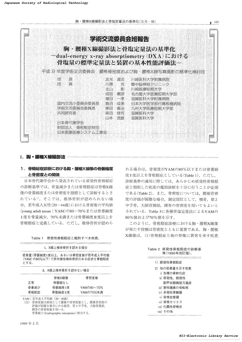
- 友光 達志 川勝 充 北山 彰 成田 憲彦 増田 一孝 鹿沼 成美 東田 善治 森田 陸司 山本 逸雄
- 出版者
- 公益社団法人 日本放射線技術学会
- 雑誌
- 日本放射線技術学会雑誌 (ISSN:03694305)
- 巻号頁・発行日
- vol.55, no.2, pp.165-187, 1999-02-20 (Released:2017-06-30)
- 参考文献数
- 15
- 被引用文献数
- 1 1


