3 0 0 0 OA インジウム化合物およびインジウム・スズ酸化物の生体影響
- 著者
- 田中 昭代
- 出版者
- 日本エアロゾル学会
- 雑誌
- エアロゾル研究 (ISSN:09122834)
- 巻号頁・発行日
- vol.20, no.3, pp.213-218, 2005 (Released:2007-01-12)
- 参考文献数
- 28
- 被引用文献数
- 1
There was little information regarding the adverse health effects to humans or animals arising from exposure to indium compounds until mid 1990. However, due to the increasingly frequent industrial use of indium such as indium-tin oxide (ITO), the potential occupational or environmental exposure to indium compounds has attracted much attention. In 2001, the first case of interstitial pneumonia caused by exposure to ITO occupationally was reported. Recent animal studies have indicated the pulmonary or testicular toxicity of indium compounds. Furthermore, carcinogenicity of InP was demonstrated by the inhalation study in rats and mice and that of InAs was suspected in the intratracheal instillation study using hamsters. It appeared that indium was toxic when released from the particles, though the physical characteristics of the particles also contribute to toxic effect. It is necessary to pay much greater attention to the human exposure of indium, indium compounds or ITO.
3 0 0 0 OA 溶接工肺―溶接ヒュームによる肺障害―
- 著者
- 吉井 千春 森本 泰夫 城戸 優光
- 出版者
- 日本エアロゾル学会
- 雑誌
- エアロゾル研究 (ISSN:09122834)
- 巻号頁・発行日
- vol.20, no.3, pp.238-242, 2005 (Released:2007-01-12)
- 参考文献数
- 18
- 被引用文献数
- 2
Welder's pneumoconiosis, which is caused by the inhalation of welding fumes, is one of the major pneumoconioses in Japan. The major component of welding fumes is iron oxide. Although welder's pneumoconiosis has been considered to be inert, recent reports revealed the possibility of developing fibrosis. In this article, we demonstrate radiological and pathological features of welder's pneumoconiosis, and also review the mechanisms of developing pulmonary fibrosis. In high-resolution CT (HRCT), typical welder's pneumoconiosis shows fine centrilobular nodules in both lung fields. In some cases, fibrotic changes may be seen in subpleural areas in both lower lung fields. Lung biopsy specimens show numerous hemosiderin-laden macrophages within alveolar spaces associated with mild to moderate interstitial fibrosis. The mechanisms of developing fibrosis can be explained by “overload phenomenon”. Namely, in cases of mild exposure to welding fumes, iron oxides and hemosiderin-laden macrophages locate within air-spaces and may be reduced in numbers by mucociliary transport system. However, in cases of massive inhalation, accumulation of iron oxides and hemosiderin-laden macrophages exceed the capacity of mucociliary transport system. As a result, they invade into the interstitium and cause interstitial inflammation or thickening and eventually pulmonary fibrosis.
3 0 0 0 超音波霧化分離の工業的応用
- 著者
- 松浦 一雄
- 出版者
- 日本エアロゾル学会
- 雑誌
- エアロゾル研究 (ISSN:09122834)
- 巻号頁・発行日
- vol.26, no.1, pp.30-35, 2011
Separation through ultrasonic atomization has advantages in industrial application over distillation, i.e., low energy consumption, unheated process and quick start of opertion. The first application of ultrasonic atomization was sake refining by utilizing the advantage of no heating, its application is now growing in various industrial processes, for example, ethanol purification, waste water treatment, micro-nanometer ice formation, sugar concentration etc. In addition, carbon dioxide emission by ultrasonic atomization separation is extremely low compared to distillation. The ultrasonic atomization separation will replace many conventional separation processes because of the superior characteristics.<br>
3 0 0 0 OA 大気中コロナ放電によるイオンの生成と発展の研究
- 著者
- 関本 奏子 高山 光男
- 出版者
- 日本エアロゾル学会
- 雑誌
- エアロゾル研究 (ISSN:09122834)
- 巻号頁・発行日
- vol.26, no.3, pp.203-213, 2011 (Released:2011-09-28)
- 参考文献数
- 49
- 被引用文献数
- 1
Ambient corona discharge has been used as an ionizer in a wide range of research and industrial fields such as environmental, analytical and aerosol sciences, and possibly even commercial electric appliances. Terminal ions produced via ion evolutions through successive ion-molecule reactions in corona discharge can readily and efficiently supply the charge for ionization in ambient air. Despite substantial progress in the application of corona discharge, the elementary processes involved in terminal ion formation and evolution are not yet well understood. It has been reported that negative ion evolution is rather complex compared to that of positive ions, and that it is difficult to regulate the reproducible formation of specific negative ion species. We have recently established an atmospheric pressure corona discharge system containing a specific corona needle that successfully leads to regular and reproducible generation of various positive and negative ions originating from ambient air. The system coupled with mass spectrometers made it possible to study the relationship between terminal ion formation and discharge conditions. In this paper, the formation and evolution mechanism of terminal ions such as H3O+, HO-, NOx- and COx- depending on the electric field strength on the needle tip and the resulting kinetic energy of electrons accelerated at the tip will be described.
3 0 0 0 OA 黄砂バイオエアロゾルに含まれる耐塩細菌群の種組成解析
- 著者
- 牧 輝弥 小林 史尚 柿川 真紀子 鈴木 振二 當房 豊 山田 丸 松木 篤 洪 天祥 長谷川 浩 岩坂 泰信
- 出版者
- 日本エアロゾル学会
- 雑誌
- エアロゾル研究 (ISSN:09122834)
- 巻号頁・発行日
- vol.25, no.1, pp.35-42, 2010-03-20 (Released:2010-03-25)
- 参考文献数
- 23
- 被引用文献数
- 2
The microbial communities transported by Asian desert dust (KOSA) events have attracted much attention as bioaerosols, because the transported microorganisms are thought to influence the downwind ecosystems in Japan. In particular, halotolerant bacteria which are known to be tolerant to atmospheric environmental stresses were investigated for clarifying the long-range transport of microorganisms by KOSA. Bioaerosol samples were collected at high altitudes within the KOSA source area (Dunhuang City, China) and the KOSA arrival area (Suzu City, Japan). The microorganisms in bioaerosol samples grew in media containing up to 15 % NaCl, suggesting that bacteria tolerant to high salinities would remain viable in the atmosphere. The PCR-DGGE (Denaturing gradient gel electrophoresis) analysis using 16S rRNA genes sequences revealed that the halobacterial communities in bioaerosol samples belonged to the members of the genera Bacillus and Staphylococcus and that some bacterial species belonging to Bacillus subtilis group were similar among the samples of both cities. Moreover, some sequences of B. subtilis group were found to be identical for the species collected at high altitudes and on the ground surfaces. This suggests that active mixing of the boundary layer transports viable halotolerant bacteria up to the free atmosphere at the KOSA source area, while down to the ground surface at the KOSA arrival areas.
3 0 0 0 OA 静電霧化微粒子水における電荷数の解析
- 著者
- 山内 俊幸 前川 哲也 瀬戸 章文 權 純博 奥山 喜久夫
- 出版者
- 日本エアロゾル学会
- 雑誌
- エアロゾル研究 (ISSN:09122834)
- 巻号頁・発行日
- vol.23, no.2, pp.108-113, 2008-06-20 (Released:2008-06-25)
- 参考文献数
- 4
We evaluate the size distribution of fine water droplets produced by electrostatic atomization using a differential mobility analyzer (DMA) and a condensation nucleus counter (CNC) . Broad size distribution with the peak of mobility diameter of around 15-20 nm is observed for the neutralized polydisperse water droplets. The number of charge is analyzed using a tandem DMA system. The water droplets are highly charged depending on the particle size. The number of maximum electric charges is 14, which is well below the Rayleigh limit.
2 0 0 0 OA 呼吸器由来の飛沫の水分蒸発過程
- 著者
- 竹川 暢之
- 出版者
- 日本エアロゾル学会
- 雑誌
- エアロゾル研究 (ISSN:09122834)
- 巻号頁・発行日
- vol.36, no.4, pp.231-236, 2021-12-20 (Released:2021-12-24)
- 参考文献数
- 28
Airborne transmission of virus-laden respiratory droplets has drawn a great deal of attention since the outbreak of COVID-19. Ambient relative humidity is the key factor controlling the evaporation of water from airborne droplets. The evaporation processes of respiratory droplets due to decreases in relative humidity are not well understood because of the complexity of the chemical compositions of the droplets. This article reviews the current understanding of the effects of relative humidity on the physicochemical and aerodynamic properties of respiratory droplets and on the infectivity of enveloped viruses.
2 0 0 0 OA 生体影響の現状
2 0 0 0 インジウム化合物およびインジウム・スズ酸化物の生体影響
There was little information regarding the adverse health effects to humans or animals arising from exposure to indium compounds until mid 1990. However, due to the increasingly frequent industrial use of indium such as indium-tin oxide (ITO), the potential occupational or environmental exposure to indium compounds has attracted much attention. In 2001, the first case of interstitial pneumonia caused by exposure to ITO occupationally was reported. Recent animal studies have indicated the pulmonary or testicular toxicity of indium compounds. Furthermore, carcinogenicity of InP was demonstrated by the inhalation study in rats and mice and that of InAs was suspected in the intratracheal instillation study using hamsters. It appeared that indium was toxic when released from the particles, though the physical characteristics of the particles also contribute to toxic effect. It is necessary to pay much greater attention to the human exposure of indium, indium compounds or ITO.
2 0 0 0 OA エアフィルタの集塵理論と静電気効果
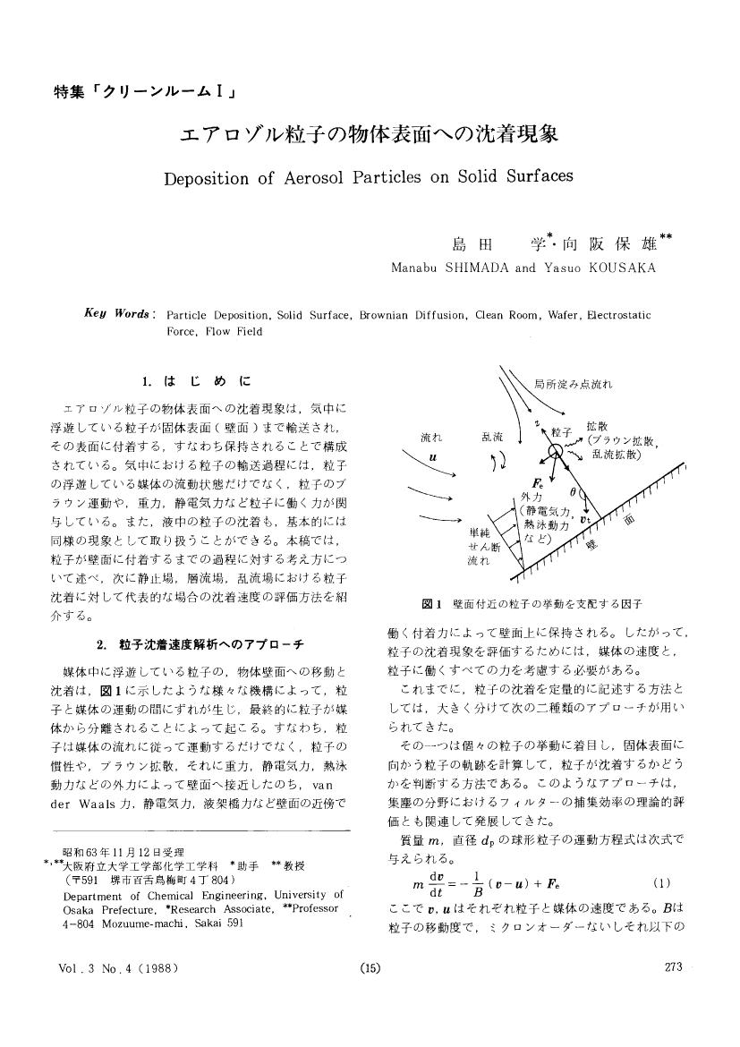
2 0 0 0 OA エアロゾル粒子の物体表面への沈着現象
2 0 0 0 OA 氷床コアに記録された気候•環境変動
- 著者
- 本山 秀明
- 出版者
- 日本エアロゾル学会
- 雑誌
- エアロゾル研究 (ISSN:09122834)
- 巻号頁・発行日
- vol.25, no.3, pp.247-255, 2010 (Released:2010-10-20)
- 参考文献数
- 21
- 被引用文献数
- 1
The accumulation rate, aerosol flux, and air temperature fluctuation can be determined from the study of ice cores drilled through ice sheets and glaciers. The aerosol which gives climate and environmental information is accumulated on the surface of ice sheet. In order to elucidate the climate and environmental changes, it is necessary to find the changes in concentration, composition, and isotope ratio of impurities in accumulated particles after the deposition on the snow surface. This study revealed that the global climate and environmental changes have occurred on various time scales in the past million years. The characteristics of aerosol particles deposited on the Antarctic ice sheet are investigated. Furthermore, the history of solar activity and associated geomagnetic fields is clarified by analyzing the cosmogenic nuclides in ice cores. An interdisciplinary study on ice cores is also carried out to elucidate the evolution mechanisms of microorganisms in the ice cores.
2 0 0 0 OA 負イオンの生理効果
- 著者
- 渡部 一郎
- 出版者
- 日本エアロゾル学会
- 雑誌
- エアロゾル研究 (ISSN:09122834)
- 巻号頁・発行日
- vol.18, no.1, pp.27-32, 2003-03-20 (Released:2007-11-27)
- 参考文献数
- 19
2 0 0 0 OA 粉体の帯電メカニズム
- 著者
- 松山 達
- 出版者
- 日本エアロゾル学会
- 雑誌
- エアロゾル研究 (ISSN:09122834)
- 巻号頁・発行日
- vol.28, no.4, pp.245-250, 2013-12-15 (Released:2014-01-07)
- 参考文献数
- 22
In this paper the mechanism of contact- or tribo-electrostatic charging of powder was briefly reviewed.Although it is often referred that ‘electrostatics is mysterious’, but the fact should be noted that the entire system of the electromagnetism, such as Coulomb’s law, is ultimately well established. After this, there are parts of knowledge on electrostatics as well understood, miss-understood sometimes, and questions leftover, and these should be separated carefully. The central question to be answered is how much amount of charge is held by each particle. On this topic, two mechanisms of charge generation and of charge fixation were reviewed and discussed in general.
- 著者
- 鈴木 基雄 登内 道彦 村山 貢司 光本 浩太郎
- 出版者
- 日本エアロゾル学会
- 雑誌
- エアロゾル研究 (ISSN:09122834)
- 巻号頁・発行日
- vol.20, no.4, pp.281-289, 2005 (Released:2007-01-12)
- 参考文献数
- 16
- 被引用文献数
- 3
Durham method has been adopted widely in Japan as the measurement method for counting the number of pollens. However, it is a tedious method for researchers because they have to count the exact number of pollens on plates using a microscope. Therefore the number of pollens has been reported only once a day from most of the observatories. In order to obtain the hourly number of pollens for further researches, automatic pollen counters have been developed.We evaluated the performance of the pollen counter KP-1000 (Kowa Co. Ltd.), which had been developed by the project of Ministry of Education, Culture, Sports, Science and Technology. In order to evaluate the capability for counting Cedar and Cypress pollen particles, we compared the number of pollens measured by KP-1000 with the number of pollens obtained by Durham method and the number of pollens caught on the outlet filter of KP-1000. Furthermore we compared the number of pollens measured by KP-1000 with the number of pollens counted by another pollen counter KH-3000 (Yamato Mfg. Co. Ltd.). The result of the comparison showed that KP-1000 could distinguish Cedar and Cypress pollens accurately.We found the high concentration of pollens in early mornings through the hourly observation of Cedar and Cypress pollens during the spring of 2005. By using the Backward Trajectory Method, we found that the high concentration phenomena of pollens were brought by the long distance transportation of pollens from Shizuoka area via Izu Peninsula and Kanagawa to Tokyo.
2 0 0 0 OA タイタンの有機物エアロゾルと生命の起源
- 著者
- 谷内 俊範 小林 憲正
- 出版者
- 日本エアロゾル学会
- 雑誌
- エアロゾル研究 (ISSN:09122834)
- 巻号頁・発行日
- vol.22, no.2, pp.113-118, 2007-06-20 (Released:2007-06-20)
- 参考文献数
- 24
- 被引用文献数
- 2
Titan is the largest satellite of Saturn, and has dense (ca. 1,500 hPa) atmosphere mainly composed of nitrogen and methane. In addition to various organic compounds, aerosol is found in the atmosphere. It is suggested that the aerosol was made of complex organic compounds, which was formed from Titan atmosphere by such energies as ultraviolet light, high-energy electrons (discharges) and cosmic rays. Voyager and Cassini missions partly revealed the nature of the aerosol. A wide variety of laboratory simulation experiments have been conducted by using a gas mixture of nitrogen and methane, and the resulting solid products are often referred as “Titan tholins”. The present paper reviews these observations and simulation experiments on the Titan aerosol, and discusses its relevance to origins of life.
2 0 0 0 OA 雲核としての有機エアロゾル研究の現状
- 著者
- 堀 雅裕
- 出版者
- 日本エアロゾル学会
- 雑誌
- エアロゾル研究 (ISSN:09122834)
- 巻号頁・発行日
- vol.18, no.4, pp.257-265, 2003-12-20 (Released:2007-10-10)
- 参考文献数
- 59
- 被引用文献数
- 2
Water soluble inorganic substances such as (NH4) 2SO4 and NaCl in aerosol particles have been considered to serve as the only cloud condensation nuclei (CCN) in the atmosphere. Recent observational studies, however, indicate that some of the organic compounds in aerosols also play an important role in the activation of CCN comparable to the inorganic particles' contribution. This article reviews recent observational and laboratory experiments conducted to measure various physico-chemical properties of organic substances that determine the high activation capability of pure organic particles and that of organic and inorganic mixtures as CCN.
2 0 0 0 OA 海洋性硫黄化合物とエアロゾル
2 0 0 0 OA シリカ粒子の肺障害関連因子 - 肺内滞留粒子量 -
2 0 0 0 OA 超音波霧化現象の可視化解析
- 著者
- 土屋 活美 林 秀哉 藤原 和久 松浦 一雄
- 出版者
- 日本エアロゾル学会
- 雑誌
- エアロゾル研究 (ISSN:09122834)
- 巻号頁・発行日
- vol.26, no.1, pp.11-17, 2011 (Released:2011-04-12)
- 参考文献数
- 19
- 被引用文献数
- 1
It has been claimed in the literature that selective ethanol separation from ethanol-water solution can be made through ultrasonic atomization. The causes of separation were explained in terms of parametric decay instability of capillary waves, accumulating acoustic energy in a highly localized surface of the capillary wave and effecting ultrasonic atomization. In this study, the atomization process is examined visually with some mechanistic view, and the dynamics of interfacial oscillations occurring along the perturbed protrusion or conical “liquid column/fountain jet” over the ultrasonic transducer are analyzed by high-speed imaging. It is found that the atomization process could be initiated by a sudden increase in surface roughness of microscale, which would be viewed as localized surface patches of two-dimensional capillary waves, often associated with contraction expansion sequence of surface to-pology. Such surface patches could bring further instability in generating a swarm of liquid droplets of microscale around the expanded phase of liquid column.


