1 0 0 0 OA ADMETローカルモデルの構築およびケミスト向けWeb GUIの開発
- 著者
- 長谷川 清 深海 隆明 大田 雅照 白鳥 康彦
- 出版者
- 公益社団法人 日本化学会・情報化学部会
- 雑誌
- ケモインフォマティクス討論会予稿集 第32回情報化学討論会 山口
- 巻号頁・発行日
- pp.O5, 2009 (Released:2009-10-22)
- 参考文献数
- 10
医薬品候補化合物としては、ターゲットに対する活性だけでなく、バランスがとれたADMET (absorption, distribution, metabolism, toxicity)特性も必要とされる。プロジェクトでの構造最適化の過程で、種々のADMET end pointsが測定され、これら特性を事前に予測できるローカルモデルがあると効率的である。われわれは、MOE descriptorsと統計パッケージRからプロジェクトごとのADMETローカルモデルを構築した。また、ケミスト用のWeb GUIを開発し、ISIS/draw, sdf fileを入力すると、予測値が自動的に出力される仕組みを構築した。
1 0 0 0 Heterocyclic Bioisostere, 基礎と応用例
- 著者
- 松岡 宏治 大田 雅照
- 出版者
- 公益社団法人日本薬学会
- 雑誌
- ファルマシア (ISSN:00148601)
- 巻号頁・発行日
- vol.46, no.3, pp.215-222, 2010-03-01
- 参考文献数
- 48
1 0 0 0 OA in silicoとin vitroの統合によるhERGチャネル阻害の回避
- 著者
- 田保 充康 長谷川 清 本多 正樹 大田 雅照
- 出版者
- 日本毒性学会
- 雑誌
- 日本トキシコロジー学会学術年会 第37回日本トキシコロジー学会学術年会
- 巻号頁・発行日
- pp.62, 2010 (Released:2010-08-18)
薬剤誘発性QT延長作用を引き起こす薬剤の多くがKチャネルの一つであるhuman ether-a-go-go related gene(hERG)チャネルを阻 害することによりQT間隔を延長させることから,hERG阻害に対する評価が創薬早期段階から実施されている。オートパッチクラン プにより多数の化合物のhERGスクリーニングが可能となり,膨大なスクリーニングの産物としてhERG阻害作用の弱い化合物を見出 すことが可能になってきた。しかしながら,薬効,薬物動態を含め薬剤としての特性を維持しながらhERG阻害の弱い化合物を効率的 に創製するためには,化合物の構造活性相関,物性情報,構造-hERG阻害相関,化合物とhERGとの相互作用などに基づいた合理的 な分子設計が必要と考えられる。特に化合物とhERGの原子レベルでの相互作用,すなわち,化合物とhERGの複合体の立体構造モデ ル情報は合理的な分子設計にとって大きなインパクトがある。hERG阻害作用を示す化合物のほとんどがhERGチャネル内部のポア領 域に結合して遮断作用を示すことが知られており,より効果的な分子設計方針を見出すためにはポア領域における結合様式を特定す ることが重要となる。hERGに対する相互作用部位として報告されているアミノ酸残基をアラニンに置換したmutant hERGに対する 化合物の阻害作用について検討し,その実験情報を考慮してhERG 3Dモデルに対する化合物のドッキングを実施することにより,化 合物の結合部位と結合様式を詳細に推定することができる。そして,この化合物/ hERG 複合体の立体構造モデル情報に基づいて, hERG阻害を回避するための合理的な構造変換アイデアの創製が可能となる。本発表では,当社の取り組みも合わせて,in vitro及び in silicoを統合したhERGチャネル阻害の回避方法について紹介する。
1 0 0 0 創薬研究の魅力を伝える(ベランダ)
- 著者
- 大田 雅照 小尾 紀行 長瀬 博 馬場 厚生 本多 利雄
- 出版者
- 公益社団法人 日本薬学会
- 雑誌
- ファルマシア (ISSN:00148601)
- 巻号頁・発行日
- vol.43, no.4, pp.301-306, 2007
- 著者
- 山本 恵子 山田 幸子 大田 雅照
- 出版者
- 公益社団法人 日本ビタミン学会
- 雑誌
- ビタミン (ISSN:0006386X)
- 巻号頁・発行日
- vol.69, no.3, pp.210-211, 1995-03-25 (Released:2018-03-17)
1 0 0 0 OA 38 活性型ビタミンDの作用発現と立体構造(口頭発表の部)
- 著者
- 山本 恵子 山田 幸子 大田 雅照
- 出版者
- 天然有機化合物討論会実行委員会
- 雑誌
- 天然有機化合物討論会講演要旨集 36 (ISSN:24331856)
- 巻号頁・発行日
- pp.290-297, 1994-09-20 (Released:2017-08-18)
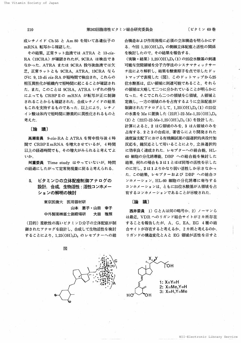
Hydroxyl groups play a major role in binding to a receptor protein. To clarify the side chain conformation of 1,25(OH)_2D_3 (1) to bind to VDR, we analyzed the mobility of the side chain of 1 and its 20-epimer (2) in terms of the spatial region accessible by the 25-hydroxyl group and the results were displayed as a dot map. The dot map indicates that each vitamin D has two major densely populated regions: A and G for 1 and EA and EG for 2. We designed six 22-substituted analogs of active vitamin D, 3-8, whose side chain hydroxyl is restricted to occupy one of the four spatial regions defined above and the analogs were classified into group A, G, EA, and EG accordingly. Syntheses of analogs, 3-8, were performed via diastereoselective conjugate addition of alkyl cuprate to steroidal (E)-and (Z)-22-en-24-ones (12 and 13) and -22-en-24-oates (16, 17, 20 and 21) as the key step. The activity of the analogs, 3-8, to bind to VDR was examined in compared with 1. It is apparent that the members of A ( 4 and 6) and EG (7) group analogs show much higher activity than the others, G(3, 5) and EA (8). The contrasting activities of the two 22-methyl analogs (3 and 4) are especially outstanding: VDR binding (3:4:1=1/50:1/3:1); differentiation of HL-60 cells (3:4:1=1/70:1:1). The results suggest that the conformation of 1,25(OH)_2D_3 (1) involved in binding to VDR and responsible for activity is the C(17,20,22,23)-anti form and the region where the 25-hydroxyl group occupies is the A.
- 著者
- 長谷川 清 深海 隆明 大田 雅照 白鳥 康彦
- 出版者
- 公益社団法人 日本化学会・情報化学部会
- 雑誌
- ケモインフォマティクス討論会予稿集
- 巻号頁・発行日
- vol.2009, pp.O5, 2009
医薬品候補化合物としては、ターゲットに対する活性だけでなく、バランスがとれたADMET (absorption, distribution, metabolism, toxicity)特性も必要とされる。プロジェクトでの構造最適化の過程で、種々のADMET end pointsが測定され、これら特性を事前に予測できるローカルモデルがあると効率的である。われわれは、MOE descriptorsと統計パッケージRからプロジェクトごとのADMETローカルモデルを構築した。また、ケミスト用のWeb GUIを開発し、ISIS/draw, sdf fileを入力すると、予測値が自動的に出力される仕組みを構築した。

