1 0 0 0 OA S状結腸捻転症を繰り返した内臓逆位症の1例
- 著者
- 田原 海 栗原 直人 松田 英士 佐々木 康裕 木村 裕子 大野 昌利 筒井 りな 松浦 芳文 飯田 修平
- 出版者
- 一般社団法人 日本消化器内視鏡学会 関東支部
- 雑誌
- Progress of Digestive Endoscopy (ISSN:13489844)
- 巻号頁・発行日
- vol.90, no.1, pp.138-139, 2017-06-09 (Released:2017-07-19)
- 参考文献数
- 4
An 81-year-old man who had been diagnosed as having situs inversus totalis and had suffered from repeated episodes of sigmoid volvulus was admitted with a history of right upper quadrant abdominal pain. Physical examination showed no evidence of peritoneal irritation. A plain radiograph of the abdomen showed a markedly dilated sigmoid colon with an inverted U-shaped appearance. Abdominal CT showed situs inversus totalis, no free air, no ascites, and a whorled appearance of the sigmoid mesentery, with dilated bowel loops. Based on these findings, the patient was diagnosed as having recurrence of sigmoid volvulus. Colonoscopy performed for repositioning showed converging mucosa signifying the distal point of the torsional obstruction, and a dilated section of the bowel with gas and feces proximal to the obstruction in the sigmoid colon. After endoscopic decompression, colonoscopy showed no evidence of mucosal ischemia. We treated this case successfully as we would have a case of sigmoid volvulus without situs inversus.
1 0 0 0 OA 若年女性に発症した胃蔓状線維粘液腫の1例
- 著者
- 森 貴裕 佐野 正弥 杉山 悟 吉原 四方 寺邑恵 里香 門馬 牧子 水上 創 中原 史雄 羽田野 敦子 藤澤 美亜 小池 潤 鈴木 孝良 松嶋 成志 鈴木 秀和
- 出版者
- 一般社団法人 日本消化器内視鏡学会 関東支部
- 雑誌
- Progress of Digestive Endoscopy (ISSN:13489844)
- 巻号頁・発行日
- vol.97, no.1, pp.73-75, 2020-12-18 (Released:2021-01-08)
- 参考文献数
- 5
A 22-year-old woman who had abdominal pain and diarrhea from 5 days ago got a CT scan in the hospital of origin and had a tumor about 5 cm in the stomach and bleeding. Upper gastrointestinal endoscopy revealed a large gastric submucosal tumor in pylorus. We considered it a malignant gastric submucosal tumor, and performed surgery, it was diagnosed as gastric plexiform fibromyxoma. Gastric plexiform fibromyxoma is a rare gastric mesenchymal tumor first reported by Takahashi et al. in 2007. Gastric plexiform fibromyxoma usually causes nonspecific symptoms of bleeding signs and is often operated on for that reason. However, surprisingly, plexiform fibromyxoma is a benign tumor with no reports of metastasis or recurrence.
1 0 0 0 OA 食道拡張バルーンを用いて除去しえた食道異物の1例
- 著者
- 濱野 徹也 光永 篤 中尾 絵美子 白戸 泉 白戸 美穂 西野 隆義
- 出版者
- 一般社団法人 日本消化器内視鏡学会 関東支部
- 雑誌
- Progress of Digestive Endoscopy (ISSN:13489844)
- 巻号頁・発行日
- vol.76, no.2, pp.56-57, 2010-06-10 (Released:2013-07-26)
- 参考文献数
- 5
症例は31歳女性。解離性障害で精神科病院入院中,27×27×12mmの円柱形異物を誤飲し当院へ紹介となった。緊急上部内視鏡施行し食道入口部に異物が嵌頓していたため,X線透視下に食道拡張バルーンを用い,バルーンとスコープで異物をはさみ,スコープごと抜去することで異物除去に成功した。食道内に隙間無く嵌頓し,鉗子での把持が困難な食道異物に対して本法は有用で,同様の状況下の様々な食道異物に対し応用可能と考えられた。
1 0 0 0 OA 大腸内視鏡検査における芍薬甘草湯(TJ-68)の腸管収縮抑制効果に関する検討
- 著者
- 相 正人 山口 武人 尾高 健夫 三橋 佳苗 宍戸 忠幸 山口 和也 税所 宏光
- 出版者
- 一般社団法人 日本消化器内視鏡学会 関東支部
- 雑誌
- Progress of Digestive Endoscopy (ISSN:13489844)
- 巻号頁・発行日
- vol.62, no.2, pp.45-49, 2003-05-31 (Released:2014-04-03)
- 参考文献数
- 7
- 被引用文献数
- 3 2
【目的】大腸内視鏡検査における芍薬甘草湯(TJ-68)の腸管収縮抑制効果を明らかにする。【対象/方法】対象は前投薬未施行で大腸内視鏡検査を行った26例。肛門から約25cmのS状結腸まで内視鏡を挿入し,全体像の把握が可能な収縮輪を規定,内視鏡との距離を保ちビデオ記録を行った。記録はTJ-68散布前3分間と,TJ-68(0.5gを微温湯50mlに溶解)を注射器で緩徐に散布した後の3分間行った。記録開始から30秒毎の計12回の画像から収縮輪内腔面積を画像解析ソフトによりpixel数として計測し,TJ-68散布前後の面積変化をグラフ化した。得られたグラフから散布前後のarea under the curve(AUC)を算出し比較検討した。【結果】TJ-68散布前のAUCは平均で41,057pixels・min,散布後は98,348 pixels・minであり,散布後有意な増大を認めた。【結論】TJ-68の大腸粘膜直接散布により明らかな腸管収縮抑制を認めた。TJ-68が大腸内視鏡検査時の収縮抑制剤となり得ることが示唆された。
1 0 0 0 OA 骨粗鬆治療薬(ビスホスフォネート製剤)内服中に重篤な食道炎・食道潰瘍をきたした1例
- 著者
- 三好 潤 大森 泰 高木 英恵 玉井 博修 川久保 博文
- 出版者
- 一般社団法人 日本消化器内視鏡学会 関東支部
- 雑誌
- Progress of Digestive Endoscopy (ISSN:13489844)
- 巻号頁・発行日
- vol.73, no.2, pp.126-127, 2008-12-10 (Released:2013-07-31)
- 参考文献数
- 2
症例は71歳,女性。ANCA関連血管炎に対するステロイド療法に伴うビスホスフォネート製剤の内服開始後47日目に心窩部痛が出現した。上部消化管内視鏡検査にて胸部中部下部食道に全周性の易出血性びらん・潰瘍が多発し,切歯列33cmに穿孔を疑う深い潰瘍を認めたが,胸部CTでは縦隔気腫・膿瘍形成を認めず保存的加療にて軽快した。本例は同剤の稀ながらも重篤な副作用である食道潰瘍が不適切な服薬状況により生じたと考えられる教訓的な症例である。
1 0 0 0 OA 潰瘍性大腸炎に伴う難治性十二指腸潰瘍の2症例
- 著者
- 久保 晴丸 森下 慎二 原口 絋 浅井 玄樹 岡崎 明佳 山川 元太 木原 俊裕 井上 雅文 松本 政雄 新村 和平
- 出版者
- 一般社団法人 日本消化器内視鏡学会 関東支部
- 雑誌
- Progress of Digestive Endoscopy (ISSN:13489844)
- 巻号頁・発行日
- vol.90, no.1, pp.108-109, 2017-06-09 (Released:2017-07-19)
- 参考文献数
- 6
A 38-year-old woman and a 29-year-old man were referred to our hospital for abdominal pain. In both cases, gastroendoscopy revealed a duodenal ulcer. H. pylori and NSAIDs (Non-Steroidal Anti-Inflammatory Drugs) intake history were negative in both cases. These duodenal ulcers were refractory even with PPI (Proton Pump Inhibitor) treatment for a few months. Colonoscopies were performed for further evaluation, and they showed vascular pattern loss and fine granular mucosal pattern. From colonoscopy and histopathological examination of the colon, we diagnosed them as ulcerative colitis. Mesalazine therapy was started, and mucosal inflammation of the duodenum and colon gradually subsided. Histopathological examinations of duodenal biopsy showed basal plasmacytosis and crypt distorsion, which is characteristic in gastroduodenitis associated with ulcerative colitis. Therefore, we diagnosed these duodenal ulcers as gastroduodenitis associated with ulcerative colitis. When we see a refractory duodenal ulcer without H. pylori infection and NSAIDs intake, we should consider the coexistence of ulcerative colitis.
1 0 0 0 OA ボノプラザン起因性collagenous colitisの1例
- 著者
- 小沼 宏徳 小沼 一郎
- 出版者
- 一般社団法人 日本消化器内視鏡学会 関東支部
- 雑誌
- Progress of Digestive Endoscopy (ISSN:13489844)
- 巻号頁・発行日
- vol.94, no.1, pp.104-106, 2019-06-07 (Released:2019-06-20)
- 参考文献数
- 9
- 被引用文献数
- 2 1
The patient was a 66-year-old woman. Approximately 2 years after she was started on oral administration of vonoprazan fumarate (potassium-competitive acid blocker [P-CAB]) for treatment of intractable reflux esophagitis, she complained of diarrheal symptoms. Symptomatic treatment was administered for the diarrhea. However, because no symptom relief was obtained, colonoscopy (CS) was performed. In the segment from the sigmoid colon to the rectum, mucosal erythema and cat scratch signs were partially observed, while histological examination revealed an approximately 30-μm collagen band directly under the mucosal epithelium. Thus, collagenous colitis (CC) was diagnosed. Oral administration of P-CAB was discontinued, and the symptoms had disappeared one month later. When CS was repeated after another 3 months, no endoscopic abnormalities were observed. Based on histological examination, the collagen band had become thinner but persisted. Most superficial epithelium had exfoliated.Though a few reports have described endoscopic monitoring of CC, ours is the first reported case of CC caused by P-CAB.
1 0 0 0 OA 縦隔炎を併発した食道胃アニサキス症の1例
- 著者
- 楠 隆昌 武井 ゆりあ 大川 修 中谷 行宏 吉野 耕平 唐鎌 優子 並木 伸 竹縄 寛 芝 祐信
- 出版者
- 一般社団法人 日本消化器内視鏡学会 関東支部
- 雑誌
- Progress of Digestive Endoscopy (ISSN:13489844)
- 巻号頁・発行日
- vol.84, no.1, pp.68-69, 2014-06-14 (Released:2014-06-21)
- 参考文献数
- 3
- 被引用文献数
- 1
Esophageal anisakiasis is a rare disease, accounting for about 0.2% of all cases of anisakiasis. A 26-year-old woman visited our hospital because of severe epigastric pain and heart burn several hours after eating pickled mackerel, which progressively worsened, and subsequently presented with fever. Contrast CT showed marked hypertrophy of the gastric wall and a significant edematous change in the lower esophagus, in addition to inflammation of the mediastinum. Upper gastrointestinal endoscopy revealed an Anisakis worm in the gastric corpus and lower esophagus. After removing it by endoscopy, her condition immediately improved. The present patient was complicated with mediastinitis. It was suggested that anisakiasis can become severe in an esophagus with a thin wall, as can be the case with the small intestine, where the worm may burrow in some cases. As in this case, we can achieve the marked amelioration of anisakiasis through the removal of worms. Thus we should perform endoscopy in the early stage. In addition, since some patients may ingest several worms of Anisakis, we should carefully check whether or not worms burrow in sites other than the stomach, where worms have already been identified.
1 0 0 0 OA コカ・コーラⓇ局注療法が奏功した胃石の一例
- 著者
- 義山 麻衣 大西 知子 前田 有紀 荒川 丈夫 飯塚 敏郎 小泉 浩一
- 出版者
- 一般社団法人 日本消化器内視鏡学会 関東支部
- 雑誌
- Progress of Digestive Endoscopy (ISSN:13489844)
- 巻号頁・発行日
- vol.97, no.1, pp.76-78, 2020-12-18 (Released:2021-01-08)
- 参考文献数
- 4
- 被引用文献数
- 2 1
A 73-year-old man was admitted with vomiting and left costal pain.He had undergone pylorus-preserving gastrectomy with B-1 reconstruction and was recently eating many persimmons. Physical examination revealed tenderness in the upper abdomen and left-side ribs. Abdominal CT showed a mass containing air in the gastroduodenal anastomosis. Esophagogastroduodenoscopy revealed a bezoar of about 10 cm in diameter in the remnant stomach. It was difficult to crush the bezoar with a grasping forceps and snare. Coca-Cola was locally injected, which softened the bezoar. We could then crush the bezoar with the grasping forceps and snare, and removed the bezoar. Oral administration or nasogastric lavage of Coca-Cola were reported as treatments for bezoars. In our case, the treatment time of a bezoar was significantly shortened by local injection of Coca-Cola and the bezoar could be treated with a small amount of Coca-Cola compared with that in previous reports.
1 0 0 0 OA オルメサルタンによる薬剤性腸炎の1例
- 著者
- 松本 悠 都築 義和 芦谷 啓吾 大庫 秀樹 市村 隆也 佐々木 淳 中元 秀友 今枝 博之
- 出版者
- 一般社団法人 日本消化器内視鏡学会 関東支部
- 雑誌
- Progress of Digestive Endoscopy (ISSN:13489844)
- 巻号頁・発行日
- vol.96, no.1, pp.151-153, 2020-06-26 (Released:2020-07-07)
- 参考文献数
- 5
Olmesartan has recently been reported as a cause of drug-induced enteropathy characterized by chronic diarrhoea and duodenal mucosal atrophy demonstrating sprue-like enteropathy. 82-year-old, male presented to our hospital because of chronic severe watery diarrhea without abdominal pain or fever. Blood examination showed mild anemia (Hb 11.2 mg/dl). Abdominal contrast-enhanced computed tomography showed mucosal edema in the large intestine. Esophagogastroduodenoscopy showed no villous atrophy in the duodenum with the possibility of pyloric gastrectomy, however, colonoscopy showed villous flattering in the terminal ileum and edematous changes in sigmoid colon. Histopathologic examination in biopsy samples from the terminal ileum and sigmoid colon showed interstitial lymphocytic infiltration. He was treated with olmesartan for hypertension at least two years before the onset of symptoms. In addition, watery diarrhea improved soon after discontinuation of olmesartan. Therefore, he was diagnosed as olmesartan-induced enteropathy. Its pathogenesis remains unclear; however, olmesartan-induced enteropathy must be included in the differential diagnosis for patients with chronic diarrhea after the intake of olmesartan.
1 0 0 0 OA 十二指腸濾胞性リンパ腫の1例
- 著者
- 大崎 篤史 芦谷 啓吾 大庫 秀樹 山岡 稔 市村 隆也 李 治平 永田 耕治 茅野 秀一 宮川 義隆 山本 啓二 中元 秀友 今枝 博之
- 出版者
- 一般社団法人 日本消化器内視鏡学会 関東支部
- 雑誌
- Progress of Digestive Endoscopy (ISSN:13489844)
- 巻号頁・発行日
- vol.87, no.1, pp.160-161, 2015-12-12 (Released:2016-01-06)
- 参考文献数
- 10
A 60-oyear-oold male visited a clinic because of gastric discomfort. This symptom was temporarily improved by a proton pump inhibitor, but it was worsened by discontinuation of the drug. He was referred to our hospital. Esophagogastroduodenoscopy showed an elevated lesion with multiple whitish small granular protrusions in the duodenal second portion, occupying two thirds of the circumference. The lesion was diagnosed to be a follicular lymphoma by histopathological examination including immunostaining of the biopsy specimens. He was admitted to our hospital. Abdominal CT scan showed no lymph node metastasis. Capsule endoscopy of the small intestine showed lymphoid follicles in the distal ileum in addition to the duodenal lesion. Bone marrow aspiration showed no invasion of lymphoma cells. This case was diagnosed as stage I according to the Lugano international conference classification. He underwent monotherapy using rituximab four times. However, the lesion did not respond. Therefore, radiotherapy was added which induced regression of the duodenal lesion. Follow-oup capsule endoscopy did not show any lesion in the distal ileum. As long-term outcome after treatment for duodenal follicular lymphoma is not known, strict observation is considered to be necessary.
1 0 0 0 OA 内視鏡検査にて確認された大腸カンジダ症の1例
- 著者
- 田崎 修平 星野 光典 福島 元彦
- 出版者
- 一般社団法人 日本消化器内視鏡学会 関東支部
- 雑誌
- Progress of Digestive Endoscopy (ISSN:13489844)
- 巻号頁・発行日
- vol.76, no.2, pp.120-121, 2010-06-10 (Released:2013-07-26)
- 参考文献数
- 4
症例は70歳男性,骨髄異形成症候群,糖尿病にて免疫能の低下した状態で下血,下痢を主訴に入院した。血清カンジダ抗原陽性,下部消化管内視鏡検査において大腸に多発性潰瘍を認め,病理組織検査にてカンジダと思われる芽胞を確認し,大腸カンジダ症と診断した。抗真菌剤の内服治療を施行したところ下痢は改善し,多発性潰瘍も縮小し,内視鏡検査にて治療前後の改善が確認できた。
1 0 0 0 OA 大腸ESDで穿孔・穿通した2例
- 著者
- 飯塚 春尚 小野里 康博 蘇原 直人 石原 弘 小川 哲史 伊藤 秀明 柿崎 暁
- 出版者
- 一般社団法人 日本消化器内視鏡学会 関東支部
- 雑誌
- Progress of Digestive Endoscopy (ISSN:13489844)
- 巻号頁・発行日
- vol.72, no.2, pp.94-95, 2008-06-15 (Released:2013-08-02)
- 参考文献数
- 3
- 被引用文献数
- 1 1
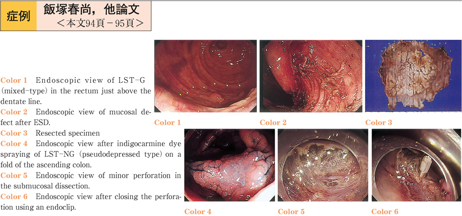
CO2送気と通常送気で大腸ESD時穿孔・穿通した症例を経験した。直腸Rbの歯状線にかかるLST-Gを通常送気で行い術後レントゲンで後腹膜気腫,皮下気腫に気づいた。術後発熱,炎症反応上昇を認めたが保存的加療し軽快。CO2送気で上行結腸LST-NGをESD中に穿孔しクリップ縫縮し,術後異常所見を認めなかった。腸管外に漏れた送気量,腸液量に違いがあるがCO2送気は通常送気と比較し大腸ESDの安全性を高める一つの方法と思われた。
1 0 0 0 OA 歴代会長
- 出版者
- 一般社団法人 日本消化器内視鏡学会 関東支部
- 雑誌
- Progress of Digestive Endoscopy (ISSN:13489844)
- 巻号頁・発行日
- vol.87, no.Supplement2, pp.s176-s361, 2015 (Released:2016-04-07)
1 0 0 0 OA 内視鏡的逆行性胆道膵管造影後に肺血栓塞栓症を発症した1例
- 著者
- 新井 絢也 戸田 信夫 黒川 憲 柴田 智華子 黒崎 滋之 船戸 和義 近藤 真由子 高木 馨 小島 健太郎 大木 正隆 関 道治 加藤 順 田川 一海
- 出版者
- 一般社団法人 日本消化器内視鏡学会 関東支部
- 雑誌
- Progress of Digestive Endoscopy (ISSN:13489844)
- 巻号頁・発行日
- vol.92, no.1, pp.148-149, 2018-06-15 (Released:2018-07-19)
- 参考文献数
- 3
A 95-year-old female was transferred to our hospital for the purpose of the treatment of cholangitis due to common bile duct stones. One day after removal of bile duct stones with endoscopic retrograde cholangiopancreatography (ERCP) , she suddenly suffered cardiopulmonary arrest. Prompt cardiopulmonary resuscitation was performed, and the spontaneous circulation and breathing resumed. Contrast-enhanced computed tomography revealed pulmonary embolisms (PE) in the bilateral pulmonary arteries. PE is known as a common but sometimes fatal complication after surgery, and prophylactic approaches including early ambulation, wearing compression stockings, intermittent pneumatic compression, and anticoagulants have been proposed based on the risk of developing PE. However, only a few reports have focused on the development of PE as a complication after invasive endoscopic procedures. In this article, we discuss the risk factors and the prophylaxis policy against PE after ERCP.
1 0 0 0 OA 90歳以上の超高齢者における大腸癌スクリーニング内視鏡検査は必要か?
- 著者
- 渡辺 一宏
- 出版者
- 一般社団法人 日本消化器内視鏡学会 関東支部
- 雑誌
- Progress of Digestive Endoscopy (ISSN:13489844)
- 巻号頁・発行日
- vol.87, no.1, pp.63-67, 2015-12-12 (Released:2016-01-06)
- 参考文献数
- 15
高齢者の大腸内視鏡検査(CS)数が増加している.当院のCSを受けた超高齢者90歳以上の患者の平均寿命と検査満足度の実態調査を行った.【対象と方法】2005年から2008年までの4年間に当院でCSを受けた当時90歳以上の78患者を対象として5年以上経過した2013年に追跡調査し,平均生存期間中央値(MST)をKaplan-Meier法を用いて対照群(当院で追跡できた年齢性別が合致した上部内視鏡検査患者群)と比較検討した.【結果】アンケート回収できた56例の検査時平均年齢91.7±1.6(90〜98)歳,MSTは3.7年で対照群と比し有意差を認めなかった.さらにグループ分けした治療群(外科/内視鏡治療)でもMSTは3.7年であった.【結論】CSを受けた90歳以上の超高齢者の平均生存期間は約4年程度であり,対照群と有意差を認めなかった.また90歳以上では有症状からでも十分なCS検査効果があると思われたが,検査を受けた患者とその家族のCS満足度は高い.今後,本邦における90歳以上のCS患者の増加が予想されるが,自覚症状のない高齢者にスクリーニングCSを何歳まで勧めるべきか平均余命を含めた議論も必要である.
1 0 0 0 OA 次亜塩素酸ナトリウム水溶液誤飲による腐食性食道炎のため食道狭窄を来した1例
- 著者
- 岡田 純卓 押本 浩一 飯田 智広 片貝 堅志 下田 隆也 増田 淳 松本 純一 荒井 泰道
- 出版者
- 一般社団法人 日本消化器内視鏡学会 関東支部
- 雑誌
- Progress of Digestive Endoscopy (ISSN:13489844)
- 巻号頁・発行日
- vol.65, no.2, pp.60-61, 2004-12-01 (Released:2014-01-28)
- 参考文献数
- 4
次亜塩素酸ナトリウム水溶液誤飲による食道炎では,多くの市販品は3%前後と低濃度である為,狭窄を生じることは少ないとされている。今回,高濃度のものによって腐食性食道炎から食道狭窄を生じることを経験したので文献的考察を加えて報告する。
1 0 0 0 OA Helicobacter pylori除菌治療に伴う血清抗体価の経時的観察
- 著者
- 松久 威史 津久井 拓
- 出版者
- 一般社団法人 日本消化器内視鏡学会 関東支部
- 雑誌
- Progress of Digestive Endoscopy (ISSN:13489844)
- 巻号頁・発行日
- vol.69, no.2, pp.31-36, 2006-12-05 (Released:2013-08-28)
- 参考文献数
- 11
- 被引用文献数
- 1
Helicobacter pylori(H. pylori)除菌治療後の血清抗H. pylori IgG抗体価低下率,抗体価陰性化までの期間とその頻度を観察した。対象は一次除菌治療に成功した519例である。血清抗体価の測定は,スマイテストELISA[ヘリコバクター]‘栄研’を使用し,H. pylori除菌前,除菌治療開始2カ月後,6カ月後,1年後の内視鏡検査時に行い,その後は年に1度の内視鏡検査時に実施した。除菌開始2カ月後の血清抗体価は32.0%低下し,3,6カ月ではそれぞれ61.4%,65.4%低下していた。H. pylori再陽性化例の存在,有意差の観点より,除菌判定には除菌6カ月後の抗体価測定が有用であることが示された。これを除菌前抗体価別にみると,400U/mL以下では60.1%,400U/mL以上800U/mL未満では67.4%,800U/mL以上では69.7%低下していた。対象症例中,抗体価陰性化例の平均期間は17.9カ月,判定保留例のそれは12.6カ月であった。両群とも抗体価の高い例(400U/mL以上)で抗体価低下率が大きかったが,抗体消失,低下には時間を要した。抗体価の累積陰性,判定保留率は除菌開始2カ月後で3.3%,除菌12,24,36,48,60,72カ月後,最長観察の77カ月後でそれぞれ14.1%,19.7%,22.0%,22.7%,23.1%,23.3%,23.5%であった。除菌成功後抗体陰性化には長期間を要した。血清抗体測定法を除菌直後の除菌判定に用いることは出来ないが,除菌成功後の経過観察に用いるのは有用と思われた。血清抗体法の長所,短所を熟知し,本法を使用すべきである。
1 0 0 0 OA 内視鏡的に除去し得た大型胃石の1例
1 0 0 0 OA 大腸内視鏡検査における炭酸ガス使用の検討
- 著者
- 乾 正幸 大和田 進 近藤 裕子 蘇原 直人 乾 純和
- 出版者
- 一般社団法人 日本消化器内視鏡学会 関東支部
- 雑誌
- Progress of Digestive Endoscopy (ISSN:13489844)
- 巻号頁・発行日
- vol.78, no.2, pp.57-60, 2011-06-10 (Released:2013-07-19)
- 参考文献数
- 5
大腸内視鏡検査に送気は不可欠であるが,空気による送気では残留空気による腹満感が出現することがある。そこで,今回スクリーニングを含む大腸内視鏡検査における炭酸ガス送気の有用性と経済性を評価することを目的とし臨床研究を行った。対象は2009年9月10日から2009年12月1日までに乾内科クリニックで施行された大腸内視鏡検査(軽処置を含む)101症例にアンケート調査を行った。この間,全例に炭酸ガス送気を用いた。また,大腸内視鏡検査1件あたりの炭酸ガスのランニングコストを,6.6kg一本の炭酸ガスボンベのコストを総検査数で除して算出した。症例の内訳は,男性49名,女性52名,年代の最頻値は60代であった。検査中及び検査後の腹満感については,楽であったと答えた群が大幅に上回った。空気送気による大腸内視鏡検査歴のある被験者で前回の検査と今回の炭酸ガス送気の検査の腹満感の比較でも楽であったとの回答が大幅に上回った。また検査中及び検査後の腹満感について統計学的検討を行ったが腹部手術歴の有無や大腸内視鏡検査歴の有無によらず腹満感は軽減されたことが示された。今回の検討期間内における大腸内視鏡検査1件あたりの炭酸ガス使用量は0.16kgであった。炭酸ガスボンベ一本6.6kgの単価は9,000円であり,1件あたりのランニングコストは227円であった。


